BGDA Practical Placenta - Cord Development: Difference between revisions
From Embryology
No edit summary |
|||
| Line 1: | Line 1: | ||
{{BGDALabPlacenta}} | {{BGDALabPlacenta}} | ||
==Placental Arteries and Vein== | ==Placental Arteries and Vein== | ||
| Line 31: | Line 20: | ||
[[File:Placental cord ultrasound 04.jpg|300px]] | [[File:Placental cord ultrasound 04.jpg|300px]] | ||
==Wharton's Jelly== | |||
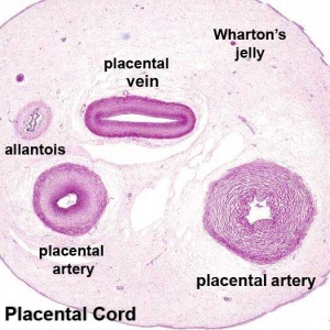
[[File:Placental_cord_cross-section.jpg|thumb|Placental cord cross-section]] | |||
* placental cord connective tissue (''substantia gelatinea funiculi umbilicalis'') | |||
* amorphous substance containing glycosaminoglycans, proteoglycans and hyaluronic acid. | |||
* cells similar to smooth muscle that allows a contractile function. | |||
* network of collagen that form canaliculi and perivascular spaces. | |||
* maintain blood flow to the fetus during placental cord compression during pregnancy or delivery. | |||
First described and named after Thomas Wharton (1614–1673) an English physician and anatomist. | |||
==Hofbauer Cells== | |||
* located the core of placental villi | |||
* macrophages with micropinocytotic activity and phagocytosis ability | |||
* possible paracrine role for early stages of placental vasculogenesis | |||
* express angiogenic growth factors (VEGF) | |||
Revision as of 19:40, 29 May 2012
| Practical 14: Implantation and Early Placentation | Villi Development | Maternal Decidua | Cord Development | Placental Functions | Diagnostic Techniques | Abnormalities |
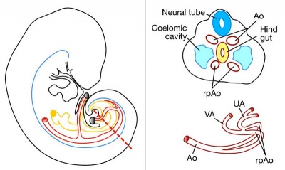
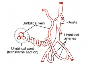
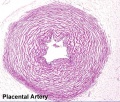
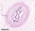
Placental Arteries and Vein
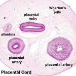
Placental Cord Histology
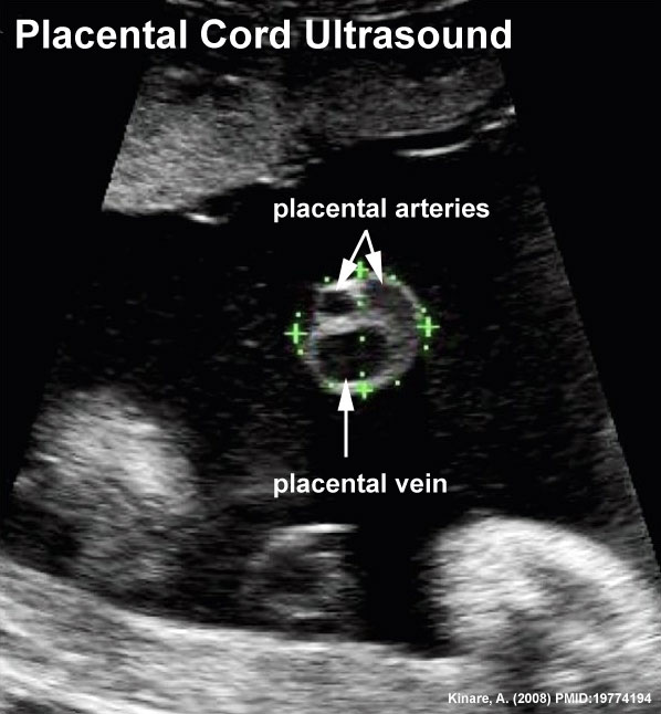
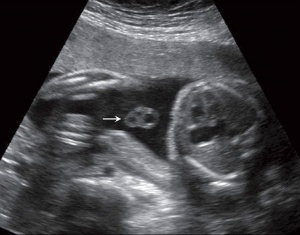
Placental Cord Ultrasound
Ultrasound image of transverse scan through the cord show the method of estimation of the cross-sectional area.
Wharton's Jelly
- placental cord connective tissue (substantia gelatinea funiculi umbilicalis)
- amorphous substance containing glycosaminoglycans, proteoglycans and hyaluronic acid.
- cells similar to smooth muscle that allows a contractile function.
- network of collagen that form canaliculi and perivascular spaces.
- maintain blood flow to the fetus during placental cord compression during pregnancy or delivery.
First described and named after Thomas Wharton (1614–1673) an English physician and anatomist.
Hofbauer Cells
- located the core of placental villi
- macrophages with micropinocytotic activity and phagocytosis ability
- possible paracrine role for early stages of placental vasculogenesis
- express angiogenic growth factors (VEGF)
Terms
| Practical 14: Implantation and Early Placentation | Villi Development | Maternal Decidua | Cord Development | Placental Functions | Diagnostic Techniques | Abnormalities |
