BGDA Practical Placenta - Cord Development: Difference between revisions
From Embryology
No edit summary |
No edit summary |
||
| Line 5: | Line 5: | ||
[[File:Placental cord vessels 02.jpg|400px]] [[File:Placental cord vessels 01.jpg|300px]] [[File:Placental cord cross-section.jpg|150px]] | [[File:Placental cord vessels 02.jpg|400px]] [[File:Placental cord vessels 01.jpg|300px]] [[File:Placental cord cross-section.jpg|150px]] | ||
==Hofbauer Cells== | |||
* located the core of placental villi | |||
* macrophages with micropinocytotic activity and phagocytosis ability | |||
* possible paracrine role for early stages of placental vasculogenesis | |||
* express angiogenic growth factors (VEGF) | |||
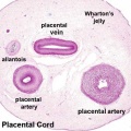
==Wharton's Jelly== | |||
[[File:Placental_cord_cross-section.jpg|thumb|Placental cord cross-section]] | |||
* placental cord connective tissue (''substantia gelatinea funiculi umbilicalis'') | |||
* amorphous substance containing glycosaminoglycans, proteoglycans and hyaluronic acid. | |||
* cells similar to smooth muscle that allows a contractile function. | |||
* network of collagen that form canaliculi and perivascular spaces. | |||
* maintain blood flow to the fetus during placental cord compression during pregnancy or delivery. | |||
First described and named after Thomas Wharton (1614–1673) an English physician and anatomist. | |||
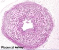
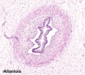
==Placental Cord Histology== | |||
<gallery> | |||
File:Placental cord cross-section.jpg|Placental cord cross-section | |||
File:Placental_vein.jpg|Placental vein | |||
File:Placental_artery.jpg|Placental artery | |||
File:Allantois.jpg|Placental allantois | |||
</gallery> | |||
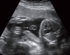
==Placental Cord Ultrasound== | |||
[[File:Placental_cord_ultrasound_03.jpg]] | |||
Ultrasound image of transverse scan through the cord show the method of estimation of the cross-sectional area. | |||
[[File:Placental cord ultrasound 04.jpg|300px]] | |||
Revision as of 19:38, 29 May 2012
| Practical 14: Implantation and Early Placentation | Villi Development | Maternal Decidua | Cord Development | Placental Functions | Diagnostic Techniques | Abnormalities |
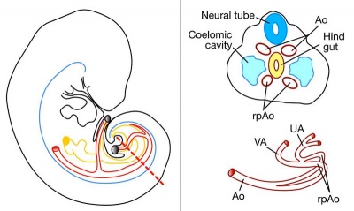
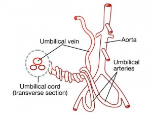
Placental Arteries and Vein
Hofbauer Cells
- located the core of placental villi
- macrophages with micropinocytotic activity and phagocytosis ability
- possible paracrine role for early stages of placental vasculogenesis
- express angiogenic growth factors (VEGF)
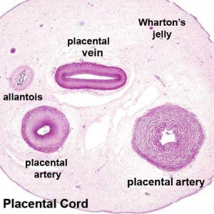
Wharton's Jelly
- placental cord connective tissue (substantia gelatinea funiculi umbilicalis)
- amorphous substance containing glycosaminoglycans, proteoglycans and hyaluronic acid.
- cells similar to smooth muscle that allows a contractile function.
- network of collagen that form canaliculi and perivascular spaces.
- maintain blood flow to the fetus during placental cord compression during pregnancy or delivery.
First described and named after Thomas Wharton (1614–1673) an English physician and anatomist.
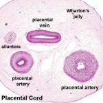
Placental Cord Histology
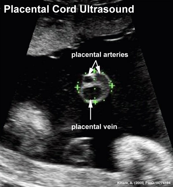
Placental Cord Ultrasound
Ultrasound image of transverse scan through the cord show the method of estimation of the cross-sectional area.
Terms
| Practical 14: Implantation and Early Placentation | Villi Development | Maternal Decidua | Cord Development | Placental Functions | Diagnostic Techniques | Abnormalities |
