2011 Group Project 1
| Note - This page is an undergraduate science embryology student group project 2011. |
Turner Syndrome
--Mark Hill 10:44, 8 September 2011 (EST) Some of the existing sub-sections have appropriate content, but there are also empty sub-sections, a total lack of referencing/citation and no glossary.
- The referencing issue needs urgent progress.
- Existing figures/table are appropriate. Table appears to be directly used from an uncited source.
- There needs to be more images in this work.
- Where is the student drawn figure?
Introduction
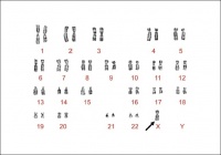
Turner syndrome (or Ullrich-Turner syndrome) is one of the most common chromosomal disorders, caused by complete or partial X monosomy in some or all cells. Approximately 1 in 2000 female live births, however the morbidity rate of spontaneous abortions is 10% and only about 1% of fetuses survive to term.
During normal fetal development, ovaries contain as many as 7 million oocytes. The oocytes gradually reduced to 400,000 during menarche and during menopause fewer than 10,000 remains. However, in Turner syndrome, the ovaries develop normally during embryogenesis but the absence of the second X chromosome leads to an accelerated loss of oocytes, which is complete by age 2 years. In this case genetically menopause occurs before menarche and the ovaries are reduced to atrophic fibrous strands, devoid of ova and follicles (streak ovaries). What is also affected is the development of somatic (nongonadal) tissues that reside on the missing/abnormal X chromosome. For example short stature is caused by a deletion of the Xp chromosome and the deletion of Xq causes gonadal dysfunction.
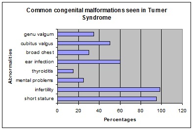
The most frequent clinical feature is short stature and gonadal dysgenesis. Other features may include coarctation of the aorta, renal anomalies, neck webbing, and lymphoedema. The affected organ systems and tissues may are effected to a lesser or greater extent amongst that are affected by turner syndrome. Each person who has turner syndrome all vary in their clinical phenotype. There has been increasing interest in this field because of the introduction in the treatment of turner syndrome with growth hormone. There has been improvement in the understanding of this condition because of the use of molecular genetic techniques. However, there still requires more work in this field. A multidisciplinary approach to treatment is important to improve the quality of life of girls with Turner Syndrome.
History
--Mark Hill 10:43, 8 September 2011 (EST) Where is this section's content?
Epidemiology
Turner Syndrome affects about 1 in 2000 live-born females. There are three types of karyotypic abnormalities but the most frequently seen is where the entire X chromosome is missing resulting in 45 X karyotype. The remaining third have structural abnormalities of the X chromosomes, and two thirds are mosaics. Whereby, the maternal X is retained in two-thirds of women and the paternal X in the remainder. The phenotype of Turner Syndrome is varies but it involves anomalies of the sex chromosome. It could be caused by the limited amount of genetic material in these abnormal chromosomes. Turner Syndrome can be transmitted from mother to daughter, and thus can it could be described as a heredity linked syndrome.
The loss of one of the sex chromosomes of turner syndrome occurs after the zygote has formed or just after the fusion of the gametes. In 70-80% of cases the retained X is from the mother. In such circumstances has lead to the different phenotypic expression of the genes present on the X chromosome depending upon if it came from the mother or father. The morphological differences from those retaining the maternal compared to retaining the paternal X is have shown to have a greater incidence of cardiovascular anomalies and neck webbing. The missing sex chromosome could be either an X or a Y. This has clinical implications because if the Y material is present there is a risk of up to 30% of gonadoblastoma developing in the dysgenetic gonads. This is because the 'gonadoblastoma locus' is on the Y chromosome which is believed to be situated on the long arm of the Y just below the centromere. Another factor affecting the phenotype in Turner syndrome is the inactivation of the abnormal X chromosomes. In a normal fetus there should be only one X chromosome that is active in each cell.
Abnormalities associated with Turner Syndrome
Etiology
--Mark Hill 10:43, 8 September 2011 (EST) Where is this section's content?
Turner Syndrome has also been linked to an increased risk of diabetes mellitus [1]
Clinical manifestations
Turner syndrome is associated with a cognitive phenotype. This includes a low IQ score, nonverbal learning disability, visuoperception deficit and attention deficity disorder. Problems with nonverbal learning involve the difficulty in mathematics. When calculating they are slow. Difficulty in both visuoperception and visuo-constructional tasks is observed when identifying and locating becomes difficult due to poor visual working memory. One other contributing factor to this is the delay in selecting visuospatial tasks. The incidence rate of attention skills and impulsivity in girls with turner syndrome is higher then children in the general population with ADHD.
Diagnostic Procedures
Diagnostic Definition
Turner syndrome is diagnosed through both evaluation of physical features and by analysis of the second sex chromosome, which in Turner syndrome is completely or partially absent (with or without cell line mosaicism). The two criteria must be met for classification of Turner syndrome, hence if physical characteristics are not present even when the cytogenetic criterion is met, the patient is not diagnosed as having Turner syndrome.[2] In addition if the homeobox gene, is not present or if there is a deletion prior to the junction between Xp22.2 and Xp22.3 the patient is also diagnosed with Turner syndrome.[3] Turner syndrome may be diagnosed prenatally and/or postnatally, as outlined below.
Prenatal Diagnosis
Postnatal Diagnosis
Diagnosing turner syndrome has been conducted prenatally and postnatally. In prenatal diagnosis is incidentally discovered during chorionic villous sampling or amniocentesis. Sex chromosome aneuploidy could occur and genetic counselling is recommended. Turner syndrome can also be detected in ultrasound findings. The increased nuchal translucency is frequently associated with turner syndrome. The presence of a cystic hygroma is another diagnosis to be most likely to be turner syndrome. Other ultrasound findings correlated to turner syndrome are coarctation of the aorta and/or left-sided cardiac defects, brachycephaly, renal anomalies, polyhydramnios, oligohydramnios, and growth retardation. Maternal serum screening of any abnormality (α-fetoprotein, hCG, inhibin A, and unconjugated estriol) may also suggest the diagnosis of turner syndrome. The diagnostic tool of turner syndrome is karyotpe confirmation and even when the prenatal diagnosis has been made by karyotype, chromosomes should be reevaluated postnatally.
In postnatal diagnosis involves the use of karyotype methods. Sufficient numbers of cells should be counted and peripheral blood karyotype is usually adequate however assessing a second tissue, such as skin, is recommended. The presence of Y chromosome material may cause the development of gonadoblastoma. Vaginal sonography along with color Doppler sonography of the gonads detects the Y chromosome materal done at regular.
Karyotyping is done when a female patient has unexplained growth failure or pubertal delay to rule out turner syndrome. Diagnosing for turner syndrome in newborn or infants is the presence of edema of the hands or feet; nuchal folds; left-sided cardiac anomalies, low hairline, low set ears, and small mandible. In childhood a decline in growth velocity and short stature and elevated levels of FSH is observed. In the newborn cubitus valgus, nail hypoplasia, hyperconvex uplifted nails, multiple pigmented nevi, characteristic facies, short fourth metacarpal, and high arched palate; extensive and chronic problems with otitis media is seen. In adolescence diagnosis of turner syndrome is the absence of breast development by 13 yr of age, pubertal arrest, or primary or secondary amenorrhea with elevated levels of FSH and unexplained short stature.
Treatment
The most common abnormality is short stature as bone age is delaged due to the lack of estrogen. This could be treated with the completion of growth hormone therapy.
For young girls use a low dose of natural estrogen to promote and maintain secondary sex characteristics. However recommended to be used when she is approaching puberty in terms of social circumstances as physical sex appearance becomes apparent among her peers.
Evaluation and Management for Turner Syndrome
| MANAGEMENT | METHOD | FEATURE |
| Orthopedic evaluation | Physical examination | Congential hip dislocation |
| Cardiac evaluation and management | Echocardiogram | BAV or COA |
| renal anatomy evaluation | Renal ultrasound | Rotational abnormalities |
| Blood pressure | Physical examination | Hypertension |
| Speech | Speech therapist | Hypertension |
| Speech | Speech therapist | Speech loss |
| Inner ear | Therapist | Hearing loss |
| Outer ear | Observation | Malformation of the outer ear |
| Middle ear | Ventilation tubes | Otitis media |
| Thyroid function | Levels of TSH | Primary hypothyroidism |
| Hearing | Karyotyping | Hearing problems |
| Plastic surgery | Elective surgery | Keloid |
| Vision | Physical examination | Ptosis |
| Orthodontic exam | Orthodontist | Dental abnormalities |
| Weight | Counselling | Obesity |
| Lyphedema | Therapy | Occur and recur at any age |
| Glucose intolerance | Growth promotion | Diabetes |
Current/future research possibilities
--Mark Hill 10:43, 8 September 2011 (EST) Where is this section's content?
Glossary
References
2011 Projects: Turner Syndrome | DiGeorge Syndrome | Klinefelter's Syndrome | Huntington's Disease | Fragile X Syndrome | Tetralogy of Fallot | Angelman Syndrome | Friedreich's Ataxia | Williams-Beuren Syndrome | Duchenne Muscular Dystrolphy | Cleft Palate and Lip


