File:Cardiac muscle EM01.jpg
Original file (1,072 × 735 pixels, file size: 231 KB, MIME type: image/jpeg)
Cardiac Muscle EM
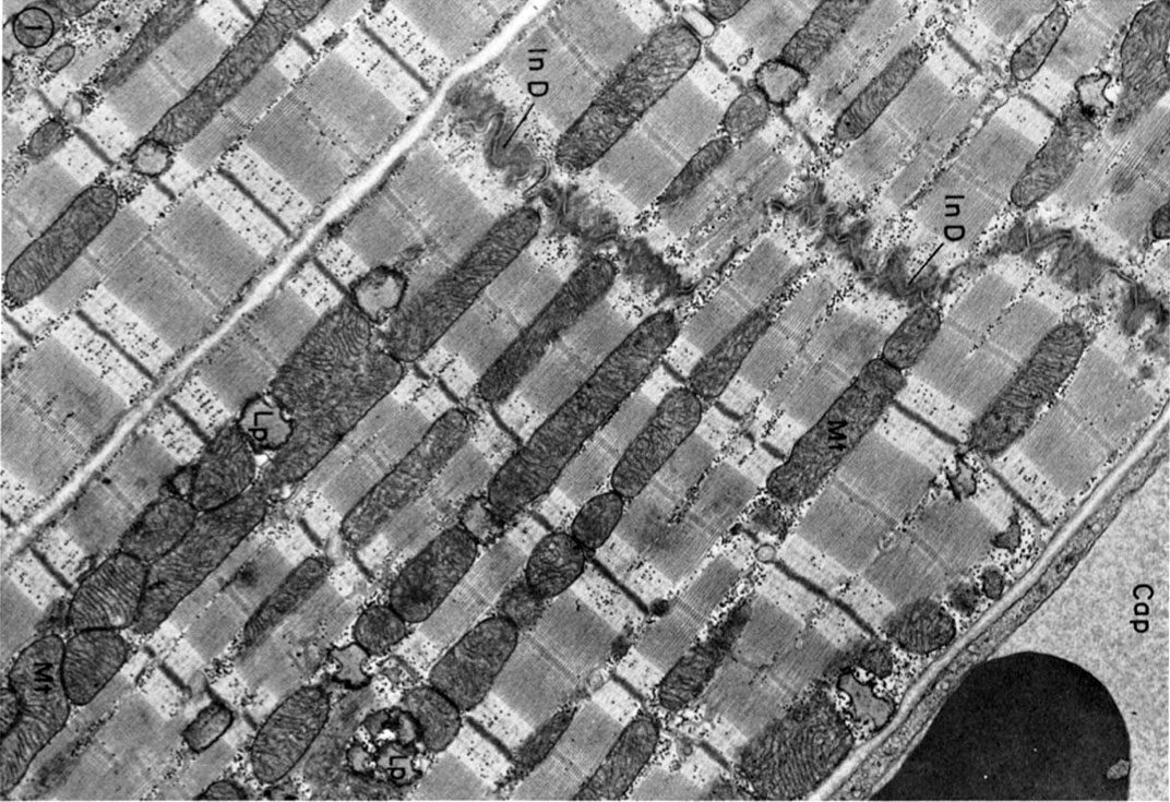
Electron micrograph of parts of three cat cardiac muscle fibers and an adjacent capillary (Cap) in longitudinal section. The two upper cells are joined end to end by a typical steplike intercalated disc (In D).Rows of mitochondria (Mt) appear to divide the contractile substance into myofibril-like units but, unlike the true myofibrils of skeletal muscle, these branch and rejoin and are quite variable in width. Lipid droplets (Lp) somewhat distorted in specimen preparation are found between the ends of the mitochondria. X 15,000.
File history
Yi efo/eka'e gwa ebo wo le nyangagi wuncin ye kamina wunga tinya nan
| Gwalagizhi | Nyangagi | Dimensions | User | Comment | |
|---|---|---|---|---|---|
| current | 12:52, 6 August 2012 | | 1,072 × 735 (231 KB) | Z8600021 (talk | contribs) | ==Cardiac Muscle EM== Electron micrograph of parts of three cat cardiac muscle fibers and an adjacent capillary (Cap) in longitudinal section. The two upper cells are joined end to end by a typical steplike intercalated disc (In D).Rows of mitochondria ( |
You cannot overwrite this file.
File usage
The following 5 pages use this file:


