File:Placenta term anatomy 01.jpg
Original file (1,200 × 600 pixels, file size: 115 KB, MIME type: image/jpeg)
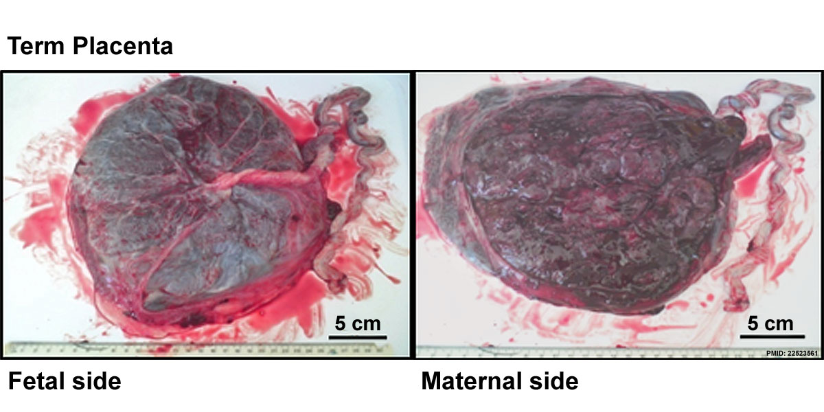
Placenta Term Anatomy
Image showing fatal and maternal sides of the placenta at term.
5 cm scale bar.
Maternal perception of reduced fetal movements is associated with altered placental structure and function
PLoS One. 2012;7(4):e34851. Epub 2012 Apr 16.
Warrander LK, Batra G, Bernatavicius G, Greenwood SL, Dutton P, Jones RL, Sibley CP, Heazell AE. Source School of Biomedicine, Manchester Academic Health Science Centre, University of Manchester, Manchester, United Kingdom.
Abstract
BACKGROUND: Maternal perception of reduced fetal movement (RFM) is associated with increased risk of stillbirth and fetal growth restriction (FGR). DFM is thought to represent fetal compensation to conserve energy due to insufficient oxygen and nutrient transfer resulting from placental insufficiency. To date there have been no studies of placental structure in cases of DFM. OBJECTIVE: To determine whether maternal perception of reduced fetal movements (RFM) is associated with abnormalities in placental structure and function. DESIGN: Placentas were collected from women with RFM after 28 weeks gestation if delivery occurred within 1 week. Women with normal movements served as a control group. Placentas were weighed and photographs taken. Microscopic structure was evaluated by immunohistochemical staining and image analysis. System A amino acid transporter activity was measured as a marker of placental function. Placentas from all pregnancies with RFM (irrespective of outcome) had greater area with signs of infarction (3.5% vs. 0.6%; p<0.01), a higher density of syncytial knots (p<0.001) and greater proliferation index (p<0.01). Villous vascularity (p<0.001), trophoblast area (p<0.01) and system A activity (p<0.01) were decreased in placentas from RFM compared to controls irrespective of outcome of pregnancy. CONCLUSIONS: This study provides evidence of abnormal placental morphology and function in women with RFM and supports the proposition of a causal association between placental insufficiency and RFM. This suggests that women presenting with RFM require further investigation to identify those with placental insufficiency.
PMID 22523561
File history
Yi efo/eka'e gwa ebo wo le nyangagi wuncin ye kamina wunga tinya nan
| Gwalagizhi | Nyangagi | Dimensions | User | Comment | |
|---|---|---|---|---|---|
| current | 14:14, 3 Haziran 2012 | | 1,200 × 600 (115 KB) | Z8600021 (talk | contribs) | ==Placenta Term Anatomy== Image showing fatal and maternal sides of the placenta at term. 5 cm scale bar. Category:Placenta ===Maternal perception of reduced fetal movements is associated with altered placental structure and function=== PLoS One. |
You cannot overwrite this file.


