File:Human spermatozoa phospholipase C zeta.jpg
Original file (1,000 × 571 pixels, file size: 119 KB, MIME type: image/jpeg)
Human Spermatozoa Phospholipase C zeta Localization
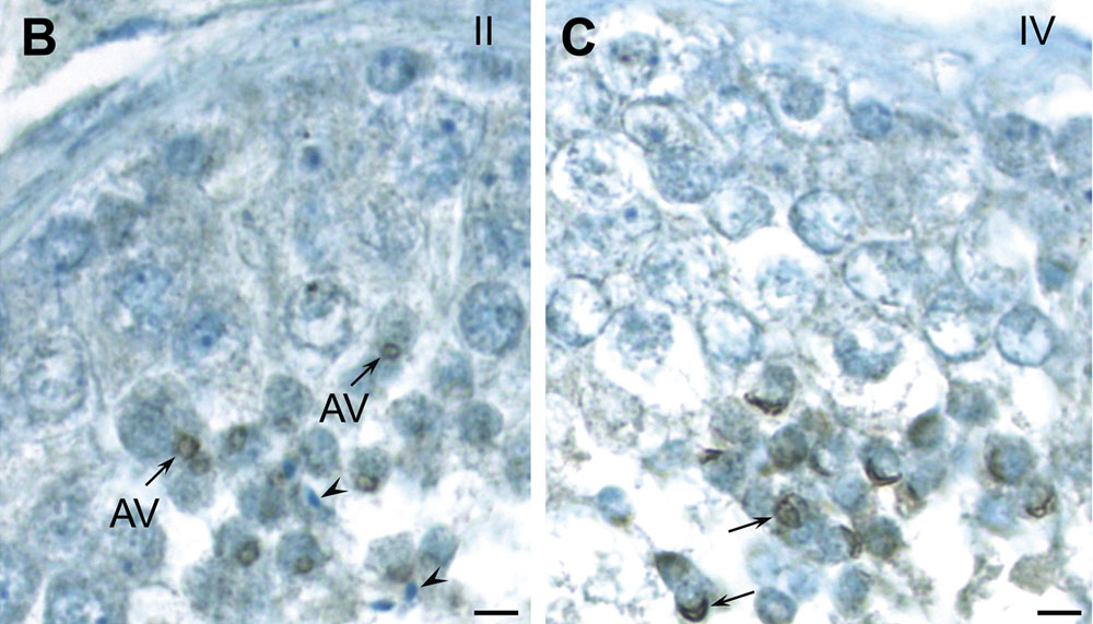
Figure 2. Developmental localization of PLCζ in mouse (A) and human (B, C) testes utilizing anti-hmPLCζ antibody. Similar results were obtained with anti-EF antibody in mouse and human and anti-hPLCζ in human sections. (A) Immunostaining originates at the beginning of acrosome formation. In stage III it appears confined to the acrosomic vesicle (AV) of step 3 spermatids as seen in detail in the inset; note that acrosomic granule is most immunoreactive. In stage VII immunostaining is intense within the acrosome, capping a portion of the nucleus (arrowheads) of the step 7 round spermatids as seen in more detail in the inset. In stages XI and XII immunostaining resides in the head of elongating spermatids in steps 11 and 12, respectively (arrows). However, by step 14 (stages II and III) the intensity of immunostaining in elongated spermatid heads has diminished significantly (arrows). (B, C) In human, there are six stages (I–VI) of the cycle of the seminiferous epithelium. As seen in B, PLCζ accumulates over the acrosomic vesicle (AV) of step 2 spermatids in stage II. Little immunoreactivity is found elsewhere in the epithelium and the elongated spermatids (arrowheads) appear unreactive. In step 4 spermatids (stage IV, see C), the fully formed acrosome is intensely labeled (arrows). Bars = 20 µm; Bars in insets = 5 µm.
http://www.ncbi.nlm.nih.gov/pubmed/22428063
http://www.plosone.org/article/info%3Adoi%2F10.1371%2Fjournal.pone.0033496
Citation: Aarabi M, Yu Y, Xu W, Tse MY, Pang SC, et al. (2012) The Testicular and Epididymal Expression Profile of PLCζ in Mouse and Human Does Not Support Its Role as a Sperm-Borne Oocyte Activating Factor. PLoS ONE 7(3): e33496. doi:10.1371/journal.pone.0033496
Copyright: © 2012 Aarabi et al. This is an open-access article distributed under the terms of the Creative Commons Attribution License, which permits unrestricted use, distribution, and reproduction in any medium, provided the original author and source are credited.
Journal.pone.0033496.g002.jpg
Panel B and C from Figure 2. Developmental localization of PLCζ in mouse (A) and human (B, C) testes utilizing anti-hmPLCζ antibody.
File history
Yi efo/eka'e gwa ebo wo le nyangagi wuncin ye kamina wunga tinya nan
| Gwalagizhi | Nyangagi | Dimensions | User | Comment | |
|---|---|---|---|---|---|
| current | 12:55, 6 May 2012 | | 1,000 × 571 (119 KB) | Z8600021 (talk | contribs) | Human Spermatozoa Phospholipase C zeta Localization== Figure 2. Developmental localization of PLCζ in mouse (A) and human (B, C) testes utilizing anti-hmPLCζ antibody. Similar results were obtained with anti-EF antibody in mouse and human and anti-hPLC |
You cannot overwrite this file.
File usage
The following page uses this file: