File:Streeter029.jpg

Original file (774 × 1,000 pixels, file size: 74 KB, MIME type: image/jpeg)
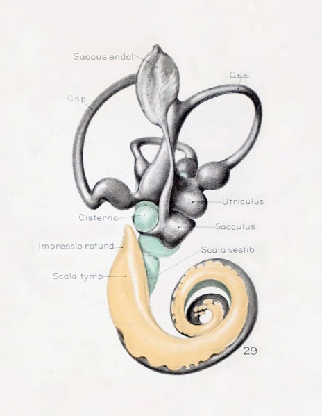
Fig. 29
Median view of same model shown in figure 28, enlarged 11.4 diameters. The scala tympani is shown in orange. The oval indentation in its proximal end corresponds to the fenestra cochlea (rotunda). This space extends along the cochlear duct about the same distance as the scala vestibuli, but the two do not commuinicate yet at any place. The peripheral border of the scala tympani is characterized by sacculations corresponding to spaces that are coalescing with the main space. The grow th of the scala is due to a coalescence of new spaces along its peripheral border rather than along its central border.
File history
Yi efo/eka'e gwa ebo wo le nyangagi wuncin ye kamina wunga tinya nan
| Gwalagizhi | Nyangagi | Dimensions | User | Comment | |
|---|---|---|---|---|---|
| current | 21:52, 22 April 2012 |  | 774 × 1,000 (74 KB) | Z8600021 (talk | contribs) | ====Fig. 29==== Median view of same model shown in figure 28, enlarged 11.4 diameters. The scala tympani is shown in orange. The oval indentation in its proximal end corresponds to the fenestra cochlea (rotunda). This space extends along the cochlear duct |
You cannot overwrite this file.
File usage
The following 2 pages use this file: