File:Adult epidermis histology 03.jpg
Adult_epidermis_histology_03.jpg (600 × 375 pixels, file size: 46 KB, MIME type: image/jpeg)
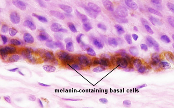
Skin Epithelium Basal Cell Layer
This is an image of the epithelial basal cell layer showing melanin (brown) that has been transferred to the keratinocyte stem cells from the nearby melanocytes.
- Integument Histology Links: Adult Skin | Epidermis and Dermis | Thin Skin Epidermis | Thick Skin Epidermis | Elastic Fibres | Basal Cell Melanin | Foundations Practical Support | Integumentary System Development | Histology Stains
Links: Histology | Histology Stains | Blue Histology images copyright Lutz Slomianka 1998-2009. The literary and artistic works on the original Blue Histology website may be reproduced, adapted, published and distributed for non-commercial purposes. See also the page Histology Stains.
Cite this page: Hill, M.A. (2024, June 22) Embryology Adult epidermis histology 03.jpg. Retrieved from https://embryology.med.unsw.edu.au/embryology/index.php/File:Adult_epidermis_histology_03.jpg
- © Dr Mark Hill 2024, UNSW Embryology ISBN: 978 0 7334 2609 4 - UNSW CRICOS Provider Code No. 00098G
File history
Yi efo/eka'e gwa ebo wo le nyangagi wuncin ye kamina wunga tinya nan
| Gwalagizhi | Nyangagi | Dimensions | User | Comment | |
|---|---|---|---|---|---|
| current | 13:26, 26 March 2012 | | 600 × 375 (46 KB) | Z8600021 (talk | contribs) |
You cannot overwrite this file.
File usage
The following 5 pages use this file: