File:Skull CT abnormal 06.jpg
From Embryology

Size of this preview: 800 × 433 pixels. Other resolution: 1,000 × 541 pixels.
Original file (1,000 × 541 pixels, file size: 85 KB, MIME type: image/jpeg)
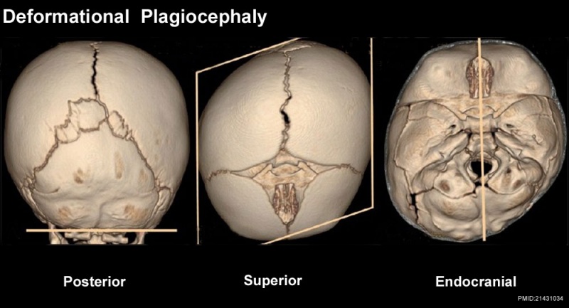
Deformational plagiocepahly
Posterior (A), superior (B) and endocranial (C) 3DCT volume rendered images. There is no identifiable synostosis. The skull shape resembles a parallelogram (B), and the posterior skull base is not abnormally tilted (A). In addition, the posterior skull base axis coincides with the anterior skull base axis (C)
http://www.ijri.org/viewimage.asp?img=IndianJRadiolImaging_2011_21_1_49_76055_f8.jpg
File history
Click on a date/time to view the file as it appeared at that time.
| Date/Time | Thumbnail | Dimensions | User | Comment | |
|---|---|---|---|---|---|
| current | 16:59, 17 March 2012 | | 1,000 × 541 (85 KB) | Z8600021 (talk | contribs) |
You cannot overwrite this file.
File usage
The following 3 pages use this file: