File:Skull CT abnormal 04.jpg
Original file (1,000 × 646 pixels, file size: 102 KB, MIME type: image/jpeg)
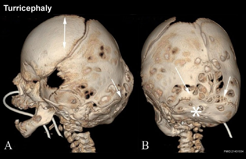
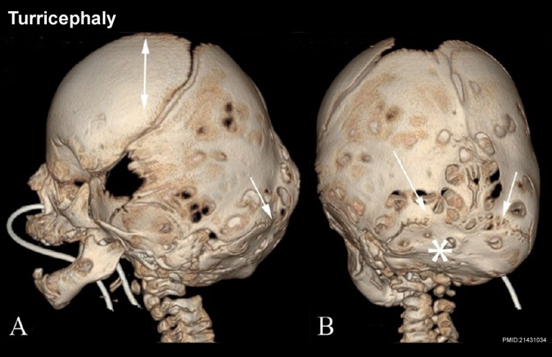
Skull Turricephaly
Turricephaly secondary to bilateral lambdoid fusion (arrows) with small, underdeveloped posterior fossa (*), and the "tall" cranium (double-headed arrow).
- A - Lateral 3DCT volume rendered image
- B - posterior 3DCT volume rendered image
Reference
<pubmed>21431034</pubmed>| PMC3056371 | Indian J Radiol Imaging.
This is an open-access article distributed under the terms of the Creative Commons Attribution License, which permits unrestricted use, distribution, and reproduction in any medium, provided the original work is properly cited.
Paritosh C Khanna © 2007 - 2012 Indian Journal of Radiology and Imaging
Attribution-NonCommercial-ShareAlike 3.0 Unported (CC BY-NC-SA 3.0)
Original file name: Figure 5 (A,B) Original figure has been modified, resized and relabelled.
http://www.ijri.org/viewimage.asp?img=IndianJRadiolImaging_2011_21_1_49_76055_f6.jpg
File history
Yi efo/eka'e gwa ebo wo le nyangagi wuncin ye kamina wunga tinya nan
| Gwalagizhi | Nyangagi | Dimensions | User | Comment | |
|---|---|---|---|---|---|
| current | 11:11, 17 March 2012 | | 1,000 × 646 (102 KB) | Z8600021 (talk | contribs) |
You cannot overwrite this file.
File usage
The following 4 pages use this file: