File:Intestine histology 002.jpg
Original file (800 × 640 pixels, file size: 130 KB, MIME type: image/jpeg)
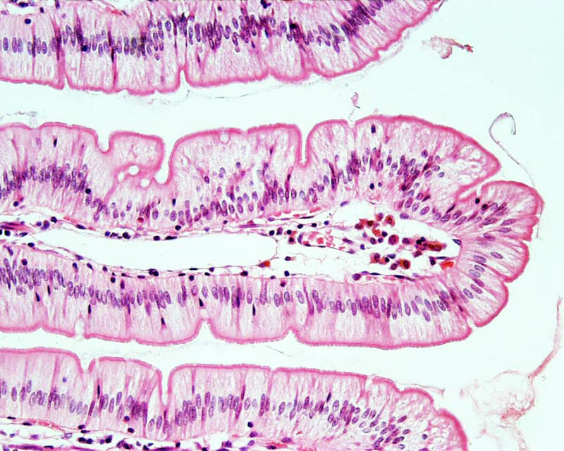
Jejunum
Histology section of Jejunum
The small intestine is divided into duodenum (25-30 cm), jejunum (about first two-fifths of the rest) and ileum. The three segments merge imperceptibly and have the same basic histological organization.
Accumulations of lymphocytes are common in the mucosa of the GIT, and they occur frequently in the small intestine. They can form very large aggregates in particular in the ileum, where they may be covered by a specialised form of epithelium which facilitates their function in the immune-defence of the body against possible pathogens in the lumen of the intestine. These specialised parts of the small intestine are called Peyer's patches,
Links: Histology | Histology Stains | Blue Histology images copyright Lutz Slomianka 1998-2009. The literary and artistic works on the original Blue Histology website may be reproduced, adapted, published and distributed for non-commercial purposes. See also the page Histology Stains.
Cite this page: Hill, M.A. (2024, June 1) Embryology Intestine histology 002.jpg. Retrieved from https://embryology.med.unsw.edu.au/embryology/index.php/File:Intestine_histology_002.jpg
- © Dr Mark Hill 2024, UNSW Embryology ISBN: 978 0 7334 2609 4 - UNSW CRICOS Provider Code No. 00098G
File history
Click on a date/time to view the file as it appeared at that time.
| Date/Time | Thumbnail | Dimensions | User | Comment | |
|---|---|---|---|---|---|
| current | 15:52, 23 February 2012 | | 800 × 640 (130 KB) | Z8600021 (talk | contribs) | |
| 15:48, 23 February 2012 |  | 800 × 640 (83 KB) | Z8600021 (talk | contribs) | ==Jejunum== Histology section of Jejunum The small intestine is divided into duodenum (25-30 cm), jejunum (about first two-fifths of the rest) and ileum. The three segments merge imperceptibly and have the same basic histological organization. Accumula |
You cannot overwrite this file.
File usage
The following 4 pages use this file: