File:T2 lymphocyte EM13.jpg

Original file (781 × 795 pixels, file size: 177 KB, MIME type: image/jpeg)
T2 lymphocyte Electron Micrograph
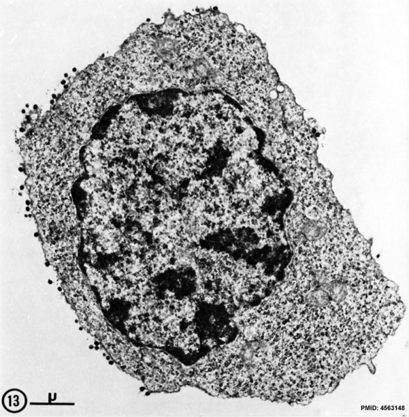
FIg. 13. Spleen cell (10 days after alloantigenic immunization with DBA/2 mastocytoma) labeled with aMSLA detected by the bridge technique with phage T4. Some of the phage heads are sectioned tangentially and therefore barely visible.
This large blast-like cell, classified as T2 lymphocyte, is mostly characterized by its very large content in polyribosomes. X 15,000.
Note this terminology may be historic.
- Lymphocyte EM Images: T and B Lymphocytes 1 TEM | T and B Lymphocytes 2 TEM | T Lymphocyte SEM | B lymphocyte 1 TEM | B lymphocyte 2 TEM | B lymphocyte 3 TEM | Plasma Cell TEM | T2 Lymphocyte 1 TEM | T2 Lymphocyte 2 TEM | lymphocyte rosettes | T lymphocyte 1 | T lymphocyte 2 | T lymphocyte 3 | T lymphocyte 4 | T lymphocyte 5 | T lymphocyte 6 | B lymphocyte | B lymphocytes TEM | Immune System Development | Blood
Reference
<pubmed>4563148</pubmed>| PMC2139311
Copyright
Rockefeller University Press - Copyright Policy This article is distributed under the terms of an Attribution–Noncommercial–Share Alike–No Mirror Sites license for the first six months after the publication date (see http://www.jcb.org/misc/terms.shtml). After six months it is available under a Creative Commons License (Attribution–Noncommercial–Share Alike 4.0 Unported license, as described at https://creativecommons.org/licenses/by-nc-sa/4.0/ ). (More? Help:Copyright Tutorial)
File history
Click on a date/time to view the file as it appeared at that time.
| Date/Time | Thumbnail | Dimensions | User | Comment | |
|---|---|---|---|---|---|
| current | 16:23, 22 February 2012 |  | 781 × 795 (177 KB) | Z8600021 (talk | contribs) | ==T2 lymphocyte Electron Micrograph== FIg. 13. Spleen cell (10 days after alloantigenic immunization with DBA/2 mastocytoma) labeled with aMSLA detected by the bridge technique with phage T4. Some of the phage heads are sectioned tangentially and theref |
You cannot overwrite this file.
File usage
The following 8 pages use this file: