File:Rat oocyte 01.jpg
Original file (1,000 × 513 pixels, file size: 55 KB, MIME type: image/jpeg)
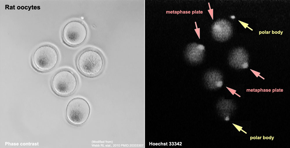
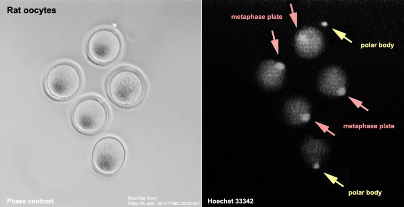
Rat Oocytes
- The same oocytes are shown for both phase contrast and fluorescence microscopy (Hoechst 33342).
- red arrows - metaphase plate is clearly visible in oocytes.
- yellow arrows - oocyte polar body.
Journal.pone.0009799.g001.png (Original image was cropped, contrast adjusted and labeled)
Reference
<pubmed>20333307 </pubmed>| PLoS One.
Citation: Webb RL, Findlay KA, Green MA, Beckett TL, Murphy MP (2010) Efficient Activation of Reconstructed Rat Embryos by Cyclin-Dependent Kinase Inhibitors. PLoS ONE 5(3): e9799. doi:10.1371/journal.pone.0009799
Copyright: © 2010 Webb et al. This is an open-access article distributed under the terms of the Creative Commons Attribution License, which permits unrestricted use, distribution, and reproduction in any medium, provided the original author and source are credited.
File history
Yi efo/eka'e gwa ebo wo le nyangagi wuncin ye kamina wunga tinya nan
| Gwalagizhi | Nyangagi | Dimensions | User | Comment | |
|---|---|---|---|---|---|
| current | 06:30, 5 November 2011 | | 1,000 × 513 (55 KB) | S8600021 (talk | contribs) | ==Rat Oocytes== * The same oocytes are shown for both phase contrast and fluorescence microscopy (Hoechst 33342). * red arrows - metaphase plate is clearly visible in oocytes. * yellow arrows - oocyte polar body. Journal.pone.0009799.g001.png (Origin |
You cannot overwrite this file.
File usage
The following page uses this file: