File:Cat oocyte zona pellucida 02.jpg
Original file (1,000 × 991 pixels, file size: 146 KB, MIME type: image/jpeg)
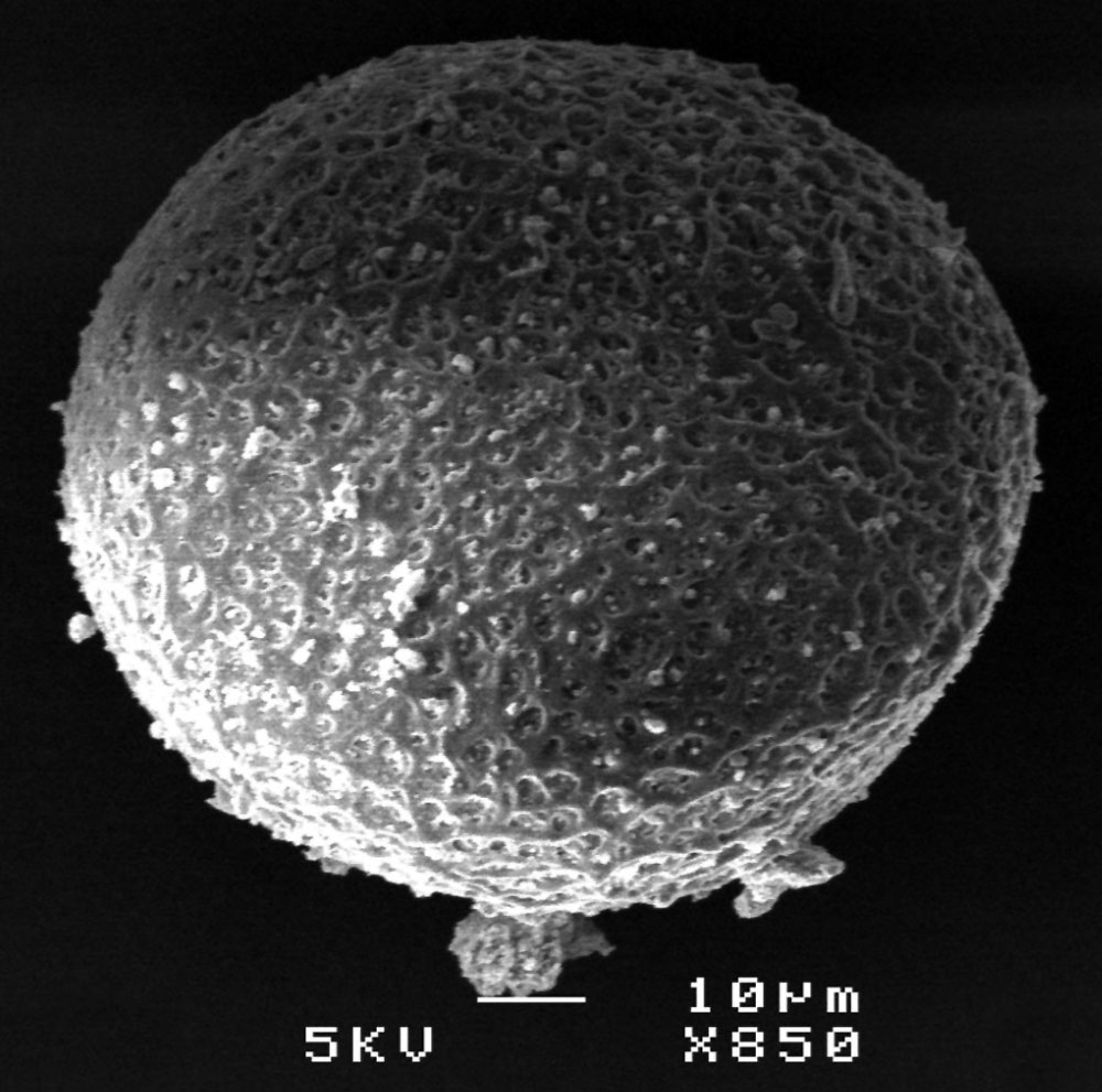
Scanning electron micrograph of the ZP of frozen-thawed feline oocytes. The size of the scale bar is 10 μm as shown in figures.
Fig. 3. 1751-0147-49-28-3-l.jpg
Reference
<pubmed>17908298</pubmed>| Acta Vet Scand.
Hermansson et al. Acta Veterinaria Scandinavica 2007 49:28 doi:10.1186/1751-0147-49-28
© 2007 Hermansson et al; licensee BioMed Central Ltd.
This is an Open Access article distributed under the terms of the Creative Commons Attribution License (http://creativecommons.org/licenses/by/2.0), which permits unrestricted use, distribution, and reproduction in any medium, provided the original work is properly cited.
File history
Yi efo/eka'e gwa ebo wo le nyangagi wuncin ye kamina wunga tinya nan
| Gwalagizhi | Nyangagi | Dimensions | User | Comment | |
|---|---|---|---|---|---|
| current | 13:34, 4 November 2011 | | 1,000 × 991 (146 KB) | S8600021 (talk | contribs) | Scanning electron micrograph of the ZP of frozen-thawed feline oocytes. The size of the scale bar is 10 μm as shown in figures. Fig. 3. 1751-0147-49-28-3-l.jpg ===Reference=== <pubmed>17908298</pubmed>| [http://www.actavetscand.com/content/49/1/28 Act |
You cannot overwrite this file.
File usage
The following 2 pages use this file:


