File:Regions of the brain.jpg

Original file (1,280 × 1,239 pixels, file size: 348 KB, MIME type: image/jpeg)
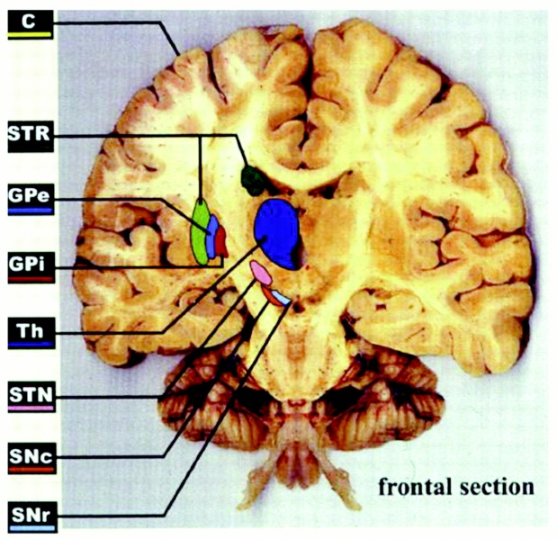
Coronal view of the brain, showing the main basal ganglia nuclei. The section is angled rostrocaudally to encounter most of the BG nuclei in a single section. C, cortex; STR, striatum; GPe, globus pallidus pars externa; GPi, globus pallidus pars interna; Th, thalamus; STN, subthalamic nucleus; SNc, substantia nigra pars compacta; SNr, substantia nigra pars reticulata.
Reference
http://physiologyonline.physiology.org/content/17/2/51.full
Organizations Requesting Permission to Reuse the Work of Others for Educational Purposes
APS grants permission for free use of our articles (in whole or in part) in educational materials provided
there is no charge or fee for those materials, and/or those materials are not directly or indirectly commercially supported. If a fee is charged or the materials are commercially supported, a fee will be assessed when permission is granted.
File history
Yi efo/eka'e gwa ebo wo le nyangagi wuncin ye kamina wunga tinya nan
| Gwalagizhi | Nyangagi | Dimensions | User | Comment | |
|---|---|---|---|---|---|
| current | 06:35, 10 October 2011 | | 1,280 × 1,239 (348 KB) | Z3290270 (talk | contribs) | Coronal view of the brain, showing the main basal ganglia nuclei. The section is angled rostrocaudally to encounter most of the BG nuclei in a single section. C, cortex; STR, striatum; GPe, globus pallidus pars externa; GPi, globus pallidus pars interna; |
You cannot overwrite this file.
File usage
The following file is a duplicate of this file (more details):
There are no pages that use this file.