Talk:2011 Group Project 11
Group 11: User:z3308965 | User:z3292953 | User:z3308968 | User:z3272325 | User:z3284061
Plagiarism
--Mark Hill 07:35, 30 September 2011 (EST) Currently all students originally assigned to each group are listed as equal authors/contributors to their project. If you have not contributed the content you had originally agreed to, nor participated in the group work process, then you should contact the course coordinator immediately and either discuss your contribution or request removal from the group author list. Remember that all student online contributions are recorded by date, time and the actual contributed content. A similar email reminder will be sent to all current students.
Please note the Universities Policy regarding Plagiarism
In particular this example:
- "Claiming credit for a proportion of work contributed to a group assessment item that is greater than that actually contributed;"
Academic Misconduct carries penalties. If a student is found guilty of academic misconduct, the penalties include warnings, remedial educative action, being failed in an assignment or excluded from the University for two years.
2011 Projects: Turner Syndrome | DiGeorge Syndrome | Klinefelter's Syndrome | Huntington's Disease | Fragile X Syndrome | Tetralogy of Fallot | Angelman Syndrome | Friedreich's Ataxia | Williams-Beuren Syndrome | Duchenne Muscular Dystrolphy | Cleft Palate and Lip
Dear Team.
Here's the Genetic Configuration and Evironmental :)
with formatting and stuff. the only 2 things missing are: an image and maybe the flow chart( same as the table in here)
Genetic Configuration and Environmental factors SECTION:
The origin of Cleft lip/ palate genetic and environmental factors arose since 1940s. A number of studies have been composed to elucidate the ambiguity behind this embryonic abnormality. It is believed that these combined factors contribute to the increase of Cleft lip/ palate incidence. Some of the genes that have been identified to have a vital role in the development of CL/CP include;
| Genetic Factors | Associated environmental Factors. |
| Transforming Growth Factor α (TGFA) | |
| Smoking | |
| Transforming Growth Factor β 3 ( TGF β3) | |
| Alcohol | |
| Muscle segment homeobox 1 (MSX1) | |
| Diatery (Folic Acid) | |
Transforming Growth Factor α (TGFA)
TGFA is regarded as one of the significant genes that have been studied extensively due to its relevance to the oral clefts. TGFA is a trans-membrane protein that is expressed at the medial edge epithelium (MEE) of fusing palatal shelves. The receptor of this gene, epidermal growth factor receptor (EGFR) is expressed on the defenerating MEE. In a study conducted on mice It was found that the deletion or alteration of this receptor contributed mostly to the facial medio-lateral defects and high occurance of cleft palate formation.
[1]
[2]
Transforming Growth Factor β 3 ( TGF β3)
Transforming Growth factor family consists of more than 30 ligands, which is believed to be involved in a numerous number of briological functions such as proliferation, differentiation, epithelium-mesenchymal transformation, and apoptosis. TFG β3 is a type of protein, known as a cytokine, which is involved in cell differentiation, embryogenesis and development. This protein was detected in the epithelial component of the vertical palatal shelf which peaks between 14-14.5 days, prior to the contact of the palatal shelves. Potential roles of the TGF β3 in cellular adhesion, and extracellular formation during the process of palate development have been demonstrated both in vivo and vitro. The alteration or mutation of this factor via smoking or other factors may result in the failure of epithelial cells during the development of palate to fuse together. Consequently, fetous is likely to develop cleft palate abnormality. [2]
[3]
Cite error: Invalid <ref> tag; name cannot be a simple integer. Use a descriptive title
Muscle segment homeobox 1 (MSX1)
Muscle segment homeobox 1 (MSX1) is a protein which is encoded by MSX1 gene. The transcripts of this protein are found in several locations such as thyrotrope-derived TSH cells, and thyrotropic tumor TtT97, pituitary cells, and most importantly expressed during embryogenesis[4]<refname="PMID10215616"><pubmed>10215616</pubmed></ref>. Due to the abundance expression sites of MSX family, it is hypothesized that these proteins might have an essential role in early development of embryo[5]. It has been discovered that MSX1, transcriptional repressor, to be significant in craniofacial, limb, odontogenesis, and nervous system development and tumor growth inhibition. Alteration in any kind including mutation of this gene may result in the formation of nonsyndromic cleft lip and/ or palate. The phenotypes of MSX1 merely depends on the locus of mutation which can affect the protein structure and function. For instance, Ser105Stop mutation was found to cause a complete deletion or absence of the MSX1 homoeodomain. This in particular impairs the development of craniofacial structures and cause clefts. [2]
Smoking:
Cleft lip and palate is considered to be one of the possible defects associated with maternal smoking. It was found that smoking cigarettes during the period of pregnancy sometimes is associated with cleft defects. The maternal glutathione s-transferase θ-1 (GSTT1) genotype, together with smoking was found to elevate the risk of cleft lip and/ or cleft palate by a ratio of 4.9. Moreover, the gene MSX1 genotypes along with maternal smoking resulted to even higher ratio of oral defects by 7.16 times. [2] [6]
Alcohol:
Several studies have shown that the teratogenic impacts of heavy alcohol exposure contributed to the incidence of cleft lip and/ or cleft palate. It was reported that about 9-18% of infacts who have experienced high alcohol exposure ended up with oral abnormalities such as cleft lip/ cleft palate[7] . However, a low level of alcohol consumption do not seem to elevate the risk of oral defects. The gene MSX1 was reported to be a major gene that could result in cleft palate or cleft lip. Furthermore, The Gene MSX1 is more likely to be altered with the consumption of four drinks or more. It was found that cytochrome Proteins may play a significant role in metabolism of endogenous morphogens in the developing fetus. Variants such as CYP2E1, or CYP1A2 were found to increase the risk of oral defects when they are altered or mutated. As a result, the structural development of the fetus is affected. [2] [8]
Folic Acid:
Maternal nutrition during pregnancy contributes highly to the normal development of foetus, especially oral and palate development. In studies conducted on mice, the development of palate was delayed due to the deficiency in folic acid supply. Moreover, insufficient dietary intake of B-complex vitamins, deficiency or excessive amount of vitamin A have been associated with high incidence of clefts formation. [9] MTHFR (5,10 methylenetetrahydrofolate reductase) is an enzyme of the metabolism of folate and homocysteine and is involved in substrate methylation and in the synthesis of nucleotides. The gene is located on chromosome 1p36.3. It was found that the absence or reduced levels of MTHFR could homocystinuria. It has been observed that a high level of homocysteine was detected in mothers with cleft babies as compared with mothers who have normal levels of MTHFR. It has been also hypothesised that variants of enzymes managing the folate metabolism contribute to the oral cleft formations. The Gene encoding encoding the MTHFR enzyme is known to have two common variants; C677T and A1298C. The link between MTHFR variants and Cleft lip and/ or palate incidences has been much less studied in the literature. [10] . [8]
Also the article of “ Complex genetics of Cleft lip and/ or cleft palate need formatting. --Maqdad Al Saif 20:21, 9 October 2011 (EST)
Hey Fleur,
This looks great! I will try and make either a table or a pie chart as the pie chart is something we haven’t done so far but depending on the level of complication of formatting I will let you know which one I can do. Are you able to prepare some text explaining the data though?
Cheers--Tahmina Lata 19:33, 9 October 2011 (EST)
hey tahmina,
ive emailed you a link to the graph and im not sure how to upload it onto here. let me know what you think/if its relevant.
--Fleur McGregor 19:27, 9 October 2011 (EST)
Sounds good. I have edited the pathophysiology without adding anything else of my own to it at this stage as I wanted to try and use most of what you had done meedo (coz I know you worked hard on it). Is this a little more understandable? I feel this section does not need a huge amount of detail as thats whats being explained in genetic configuration and development. agreed?
Pathophysiology SECTION:
The embryonic development of the upper lip and nose requires a precise genetic sequence of events. This cranio-facial development pathway is a very complex process and therefore there are several points during development at which cleft lip or palate can occur. During the third and eighth week, five major facial prominences are fused together. The lips then develop between the third and seventh week followed by the palate between the fifth and twelfth week. Due to this intricate process, multiple genetic and environmental factors will vary in their affect on the type and severity of cleft lip and palate and lead to the malfunction of the various tissues involved.
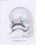
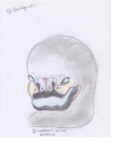
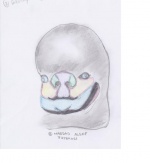
[Insert one of the Drawings from the 3 below]
The maxillary, medial nasal and lateral prominences fuse together via a combination of processes including apoptosis, epithelial bridging and subepithelial-mesenchymal penetration [insert photo for the convergence]. Cleft lip and cleft palate result when these tissues fail to fuse. Some research suggests that this abnormality is secondary to a defect of mesenchymal growth or epithelial bridging. There is evidence that intracellular signaling pathways and a wide range of errors in genetic programming may interfere with the fusion of median nasal and maxillary prominences. Consequently, the blood supply and musculature are often compromised and lead to malfunction of the lip and palate.
For example, in a unilateral cleft lip, the deep fibres of the orbicularis oris muscle are interrupted by the presence of the cleft and insert into the nasal base (the side of the defect), however, in a normal infant, these muscles will form a concentric muscle around the mouth. Furthermore, the superficial layer of the orbicularis oris muscle changes direction and forms superiorly, parallel to the edges of the cleft and inserts inferior to the columella. This can be seen when infants smile as the base of the nose would spread out laterally.
Usually, fusion of both lateral palatal shelves as well as nasal septum in the anterior posterior direction from the incisive foreman (key landmark in the bony palate-glossary) to the uvula is essential for the palate development to progress [ADD photo]. The occurrence of cleft palate is often linked with a split uvula. Functional impairment and secondary complications often occur with the gap between both the nasal and oral cavities including problems with speaking, ear infections/hearing loss, aesthetic problems, dental anomalies, psychosocial problems and hyper-nasal voice resonance due to the leakage of air from the nasal cavity.
--Fleur McGregor 19:13, 9 October 2011 (EST)
Dear Team,
It is great to see that we are all on board now. I haven't heard from Beth yet but I think the page is going to be a knockout considering the progress that has been achieved so far. Now to summarise this is what is happening:
Tonight:
Meedo-completing Genetic and environmental influences on clefting(including one image for this secion. Also writing Aetiology as it is more relating to his section.
Rahul-completing Development(adding an intro paragraph to development) and adding any other info he sees fit. Also completing current research.
Fleur-rewriting Pathophysiology and adding a graph for epidemiology including a brief discussion about the prevalence of clefting in Australia.
Meedo, the images for Pathophysio are great! Well done. Just wondering if you are able to darken the outlines of these images so it is a better resolution when they are uploaded? Having more than one student drawn image really makes the page look authentic and gives it a character. so I am quite keen on adding as many of them as we can obviously as long as it is relevant to Fluer's text on the topic.
We are hoping to have everything uploaded tonight so we can ask Dr Hill for an opinion tomorrow.
Tomorrow
- First I am going to try to get help from Dr Hill so we can get the page back!
- Then we(Rahul and I) will try to make an appointment with Dr Hill and get him to help us with the technical problems in terms of uploading images.
- Then once the page is up we will look into editing and formatting and most importantly completing the glossary.
So that is the plan at this stage. We will be in touch and please let me if you require any alterations to these.--Tahmina Lata 18:46, 9 October 2011 (EST)
http://nursingcrib.com/nursing-notes-reviewer/maternal-child-health/cleft-lip-and-palate/
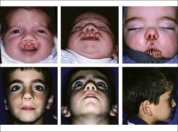
an awesome picture on this website...the one with 9 squares. not sure if we need anymore images though??
--Fleur McGregor 18:01, 9 October 2011 (EST)
Sorry to confuse you. That is what we have agreed Rahul-keep development as a standalone section, and then go onto a brief section of aetiology and then go onto genetic config- since there's more of a link between aetiology and genetic config.
This is to be added to epidemiology so please don't touch at this stage.
EPIDEMIOLOGY: Cleft lip or palate affects one in every 700 babies born in Australia. (http://www.cleftpals.org.au/about.asp) The national rate of cleft palate (1982-1992) was 4.8 - 6/10,000 births. This represents 1,530 infants, 5.5% were stillborn and 11.5% live born died during neonatal period. Cleft palate was slightly more common in twin births than singleton. The national rate (1982-1992) for cleft lip was 8.1 - 9.9 /10,000 births. Of 2,465 infants, 6.2% were stillborn and 7.8% live born died during neonatal period. The rate for cleft lip was similar in both singleton and twin births. (http://embryology.med.unsw.edu.au/embryology/index.php?title=Lecture_-_Head_Development#Cleft_Lip_and_Palate) Australian Deaths from Cleft Lip and/or Palate: 2000: 3 males, 0 females 2007: 1 male, 3 females 2008: 4 males, 0 females 2009: 3 males, 0 females http://www.abs.gov.au/ausstats/abs@.nsf/Products/CA68D0ABFECFDCF4CA2578920015B9AB?opendocument)
--Fleur McGregor 17:43, 9 October 2011 (EST)
Fleur- i thought we were gonna keep development as a standalone section, and then go onto a brief section of aetiology and then go onto genetic config- since there's more of a link between aetiology and genetic config. mm. or i thought that was what's going on. could tahmina clarify? --Rahul Mohan 17:38, 9 October 2011 (EST)
Hey rahul,
I was going to expand on it a bit more if we didnt combine sections however there is only so much i can say without doubling up on development hence the idea of combining. Im happy to keep them separate if thats best too?
there were the dot points then i pasted that other stuff further up the discussion under aetiology as well. does this clarify things?
so i just spoke to tahmina...just clarifying that meedo you are taking on aetiology now and i am working on improving pathophysiology text that you have written?
also meedo, im happy with the image so far but i will re-evaluate when its in its correct format on the oage. But at this stage, no change is required :)
--Fleur McGregor 17:22, 9 October 2011 (EST)
Too bad, we can't see the page. I think the aetiology and development combined could look good. But I reckon combining them when they are completely different titles, may not a great idea now! since we can't see both sections together. I don't know. I haven't read the page completely yet. Once we get it back, we can decide. For now, My vote is neutral :)
Thanks Fleur, IS there anything in particular all of you want me to add for the image?
Aetiology looks fantastic from the work.
I reckon final touches when we check the page as a whole, would make it phenomenoal.
I will help in other sections once I'm done with the Genetic :)
Cheers Team.
--Maqdad Al Saif 17:10, 9 October 2011 (EST)
alright- so we're gonna combine aetiology and dev. the points you listed below-is that all that aetiology is gonna be? if so- i'll put both together into one section. intro needs to be changed too. fleur could you clarify this? danke! our page is definitely coming together well. at this rate, its gonna be one of the best. looking forward.
--Rahul Mohan 16:52, 9 October 2011 (EST)
tahmina...i think that this might be some of the epidemiology stuff that was on the page??
Like many other congenital conditions prevalence of associated anomalies with cleft palate or lip has different rate and patterns of occurrence among different populations. At birth white population has higher prevalence than black population. Oral cleft in black population has been reported to have clubfoot and polydactyl as an associated anomaly as opposed to other ethnic population.[11].The reason for this is supposedly because these two anomalies are more common in the black population in general.[12] Cleft lip is more prevalent in males while cleft palate is more prevalent in females, the ratio between females and males being 60:40. From research, neither age nor parity appear to be directly related is both abnormalities. There is a higher occurance in Japanese people and lower occurance in Negro populations. The frequency of this birth defect is about 1 in 2,000 newborns in white populations.
--Fleur McGregor 16:51, 9 October 2011 (EST)
More aetiology stuff...
Genetic factors contributing to cleft lip and cleft palate formation have been identified for some cases, however, knowledge about genetic factors that contribute to the more common isolated cases of cleft lip/palate is still uncertain. Many clefts run in families, even though in some cases there does not seem to be an identifiable syndrome present, possibly because of the current incomplete genetic understanding of midfacial development. In some cases, cleft palate is a secondary issue caused by some syndromes or chromosome disorders. For example, Patau syndrome, Hardikar syndrome, Stickler’s syndrome, Treacher Collins syndrome and Loey-Dietz syndrome. The development of the face is coordinated by complex morphogenetic events and rapid proliferative expansion, and is thus highly susceptible to environmental and genetic factors. During the first six to eight weeks of pregnancy, the shape of the embryo's head is formed. During this time, primitive tissues form and if these tissues fail to fuse, a gap appears. This may happen in one, several or all locations. These back portions are called palatal shelves, which grow towards each other until they fuse in the middle.This process is very vulnerable to multiple toxic substances, environmental pollutants, and nutritional imbalance. The biologic mechanisms of mutual recognition of the two cabinets, and the way they are glued together, are quite complex and obscure despite intensive scientific research.[20]
--Fleur McGregor 16:49, 9 October 2011 (EST)
Hey guys!
that is devastating about our page...fingers crossed Dr Hill can recover it tomorrow!!
apart from that dilemma...i can see you boys really worked hard last night so well done. from yesterday afternoon, alot of work as been done and we are really coming together as a team! so congrats to all of you!
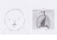
meedo-the drawings look fantastic!!! im really impressed. I vote for drawing number 1 for the intro but i think they are all excellent.
i am currently working on a few things (obviously limited until the page is up again)...I am doing a bit of epidemiology and going to help with pathophysiology to get it polished. Tahmina and I decided that it would be best to combine aetiology and development to make a more complete section as the information is similar. Is everyone ok with that? So Rahul, that intro you are working on for development you can make it an aetiology focus to intro development...here is some of the aetiology stuff i had in dot point (i didnt save a final copy)
• Cleft lip and cleft palate often occur together however have different aetiologies • Many factors cause this • Possible interaction of indirect genetic factors with environmental factors, or perhaps environmental factors alone • Cleft palate: 1 week delay in palatal shelve elevation in females, which occurs at week 8 as compared to week 7 in males o Errors in development include inadequate growth of the palatine shelves, failure of the shelves to elevate at the correct time, an excessive wide head, failure of the shelves to fuse and secondary rupture after fusion o Some mutations are linked to cleft palate and cleft lip • Cleft lip: underdevelopment of the mesenchyme of the maxillary prominence and medial nasal process o Pathogenic factors include inadequate migration or proliferation of neural crest cell ectomesenchyme, and excessive cell death during the developmental formation of the maxillary prominence and nasal placode o Some common drugs which can cause the defect include phenytoin, Dilantin, vitamin A, and some vitamin A analogs (Wong FK, 2004) • Alcohol, hypoxia, and dietary deficiencies (ie, folic acid deficiency) also have been implicated. Polygenic clefting describes a number of different genes with additive effects that result in a genetic predisposition for clefting. http://emedicine.medscape.com/article/1280866-overview
hopefully thats helpful for you Rahul.
anyways, unfortunately i didnt get to see alot of the content uploaded last night but am looking forward to seeing our page tomorrow. I think it will look great! Weve certainly come along way!!
--Fleur McGregor 16:41, 9 October 2011 (EST)
Hello Guys,
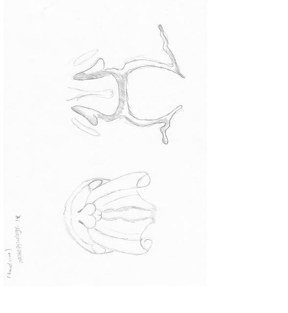
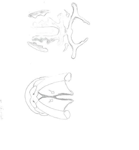
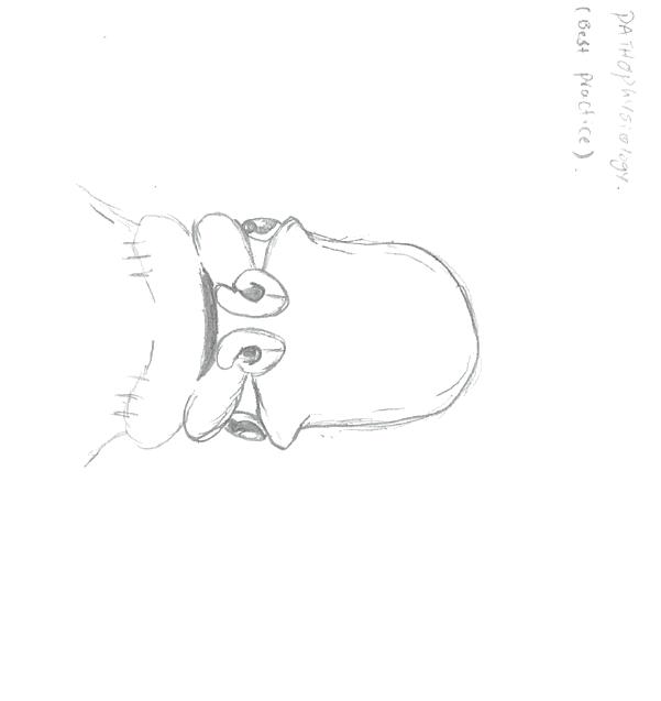
As per our previous conversations, Since the pathophysiology is standing out much within the group standards, and you would like to make big modifications. I've uploaded the prior work of mine along with the images I've drawn( if you would like me to label them and add colours) please let me know. As I'm not sure whether you need them or not. But in general this is what they will look like. Also, the Ultrasound Images that has been uploaded could be handy.
Note: if you can, try not to delete the whole section. I did a lot of work on it.
Pathophysiology SECTION:
The Development of upper lip and nose in embryos involves a series of genetically programmed procedures. This includes the fusion of five major facial prominences, that occur in gestation period between the third and eighth week. The palate develops within the fifth and twelfth week while lip develops between the third and seventh week. [Insert one of the Drawings from the 3 below]
The cranio-facial development pathway is a very complex process. Since the several points of development at which “Clefting” might occur is based on the condition and the wide range of its phonotypical expression.
In the case of Cleft Lip or palate, there’s a converge of maxillary, medial nasal and lateral prominences via a combination of few processes that include apoptosis ”programmed cell death”, epithelial bridging and subepithelial-mesenchymal penetration[insert photo for the convergence]. Cleft lip or palate are found to be secondary to a defect of mesenchymal growth or the epithelial bridging. There has been evidence stating that intracellular signalling pathways and a wide range of genetic loci may play a potential role in elucidating this abnormality. These possible proposals could cause the fusion of median nasal and maxillary prominences to be disturbed. Consequently, the blood supply in bilateral cleft lip and/or palate, especially the arterial network and musculature of the lateral elements seems to be similar to the lateral segment of the unilateral deformity. The blood vessels and muscle fibers run along the margins of the piriform aperture and prolabial segment toward the and run columella , where they anastomose with nearby vessels(pre-maxillary vessels.
In the unilateral cleft lip: the deep fibres of the orbicularis oris muscle are disturbed by the presence of the cleft and insert on the side of the defect (nasal base) as compared to the normal infant, these muscles will make their way around the mouth. Furthermore, the superficial component of the orbicularis oris changes direction and head superiorly, parallel to the edges of the cleft and insert inferior to the columella. This can be seen when infants smile as the base of the nose would splay laterally.
In cleft palate: Clefts of the palate are seem to be associated with bony and soft-tissue abnormalities. Usually, fusion of both lateral palatal shelves as well as nasal septum in the anterior posterior direction from incisive foreman ( key landmark in the bony palate ) to the uvula is essential for the palate development to progress[ADD photo]. As mentioned previously, the cleft palate is often formed the palatal development is disturbed between the 5th and 12th week of gestation. The occurrence of Cleft palate is often associated with a split uvula. Some issues are associated with the gap between both the nasal and oral cavities. These includes problems in speaking ear infections/hearing loss, aesthetic problem, dental anomalies, psychosocial problems and hyper-nasal voice resonance due to the leakage of air from the nasal cavity
[13]
[14]
[15]
Here are the images for Pathophysiology section.
--Maqdad Al Saif 16:08, 9 October 2011 (EST)
roger that. workin on it. updated dev should be up within the hour. wanna include a bit more info.
--Rahul Mohan 15:22, 9 October 2011 (EST)
Hey Rahul, can you also add a quick introductory paragraph to the development? Like a summary explaining what is it exactly that causes cleft lip and palate and then ease into the developmental staging discussion? --Tahmina Lata 14:51, 9 October 2011 (EST)
Rahul, I thought the gif and the jpeg uploading is exactly the same?? Have you tried to upload it using the same coding as the image uploading?--Tahmina Lata 10:10, 9 October 2011 (EST)
oh- and the gif image- i'm going to edit again to include more labels of the different processes, to further ease the understanding of the reader. figured out how to include the initial image provided by beth as well. so all in all, under development, we'll have the animation, the time line and the image of the development of the palate.
i'm going to need our text back to start editing the various sections. there's much to be done still. and can someone please help me out by telling me how to have a gif image show on the wiki? i can't seem to find any examples so far.
--Rahul Mohan 04:32, 9 October 2011 (EST)
Development
The palate and the lips are developed from 3 of the 5 prominences that surround the stomodeum of the fetus: the frontonasal prominence, the right maxillary prominence and the left maxillary prominence.
At stage 14 of the Carnegie staging system, the surface ectoderm of the ventrolateral part of the frontonasal prominence undergoes thickening to form the nasal placodes. The remaining portions of the frontonasal processes then grow and bulge around these placodes to form the nasal pits and the nasal processes. The mesenchyme of the maxillary process continues to grow, pushing the nasal pits medially. The upper lip consists of the maxillary processes and both the medial and lateral nasal processes- which have commenced fusion.
At stage 15- the maxillary and medial nasal processes undergo rapid growth and push the lateral nasal processes rostrally. The medial nasal process and the maxillary process also undergo fusion distally. Active fusion takes place between the lateral nasal and medial nasal processes- commencing initially at the posterior part of the nasal pits, and proceeding henceforth in an anterior direction.
From stage 16 to 18, epithelial fusion continues between the maxillary, and the 2 nasal processes. The maxillary processes continue fast growth and this results in the protrusion of the nasal pits and medial nasal processes mediofrontally. The ongoing growth and confluence of the medial nasal and maxillary mesenchyme result in the groove located in between the medial nasal processes leveling out and becoming smooth. Fusion between the medial and lateral nasal processes takes place, resulting in nasal pits to be transformed into nasal chambers and ducts.
Stage 18 heralds the connection of the nostrils to the posterior oral cavity. The medial nasal and maxillary processes also undergo fusion, resulting in the remodeling of the lower edge of the nostrils. By stage 19, the epithelial seams and the mesenchymal confluence between the medial nasal and maxillary processes disintegrate, thereby completing the formation of the upper lip.
During this stage, at about day 45, bilateral palatal shalves arise from the maxillary processes. Initially, these processes grown vertically down the side of the tongue, but then subsequently elevate to a horizontal position about the dorsal surface of the tongue. A midline epithelial seam, which undergoes degeneration to form a continuous mesenchyme, is then formed as the medial edge of the palatal shelves fuse. In the mesenchyme of the hard palate, the osteogenic blastemata for the palatal processes of the maxillary and palantine bones differentiate, while in the soft palate, patterned myogenic blastemata develop.
Palatal development usually ends (trying to find out an approximation that fits here.)
(this is just the section on the development of the palate and the upper li. still to come: information about how clefting in the palate can happen due to a disturbance in any of these processes during the palate development. trying to find more information on where and what causes clefting in the upper lip. Tahmina- could you edit the bar timeline thingum to indicate that palate development is from stage 14 to stage 18 <including> that should be sometime in mid week 5 to almost the end of week 7)
off to bed. really need a snooze. more tomorrow.
--Rahul Mohan 03:59, 9 October 2011 (EST) --Rahul Mohan 04:26, 9 October 2011 (EST)
hey guys- looks like this page is still not back yet. anw, i took a look at Dr Hill's work- he's used actual movies, whereas our pic is a gif image. anyone has any idea? working on dev still. --Rahul Mohan 02:56, 9 October 2011 (EST)
People,
Just so you know, I should have my genetic configuration done by Sunday. As I'm doing a big modification. Also, Pathophysiology I'll revise it again. :)
forgot to mention, the image of the Introduction is not finalized yet, I will modify it a bit but will need all of your comments and feedback.
Nice progress team--> I saw the development changes before the page disappears. I start to like the page more...
Keep it up team.
--Maqdad Al Saif 01:12, 9 October 2011 (EST)
Yea, I checked that!!!!! It seems to be done after 12:00AM. I had a look at other pages, but their content is still there!!!!
We shall contact Dr.Hill tomorrow in regards to this issue.
--Maqdad Al Saif 00:53, 9 October 2011 (EST)
Everyone!!!!
Our page is gone...I emailed Dr Hill and also sent him a screen shot. I dont think he will reply before Monday. I am hoping he will check is email tomorrow. This is so bad! What do we do now!!!!!
--Tahmina Lata 00:28, 9 October 2011 (EST)
Guyss,
You might wanna check the page... because everything has disappeared. the only thing left is the introduction.
So what happens now?
Rahul- you may wanna check some of Dr.Hill Tutorials or some of his lectures. consider the format of animation.
--Maqdad Al Saif 00:10, 9 October 2011 (EST)
guys, i need help. i've put together the gif image- but it doesnt seem to show. any idea how to embed it into the wiki? (http://embryology.med.unsw.edu.au/embryology/index.php?title=File:Palate_and_lip_development_animation.gif)
--Rahul Mohan 23:32, 8 October 2011 (EST)
Hello People,
Sorry the uploading took longer than what I expected.
Anyways, here are all the images.
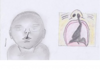
Introduction:
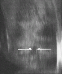
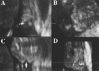
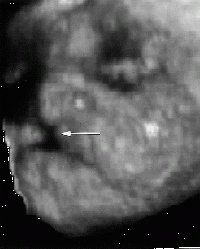
Ultrasound:
Development:
why there are 4 images for introduction, it is for you guys to decide which one will look better as an intro image. So choose 1.
Sorry but it takes a lot of time to complete the drawings and make them as presentable as possible.
The ultrasound. I will upload them on the page. You are free to move them around.
Development- Rahul, I think now, you are free to make the animation. Also, I have emailed you a higher resolution ones, since the website is a little slow in uploading images greater than 1MB.
Now, I think I have completed my duty in the drawing part. But do not forget to vote on which image for the introduction.
Is everyone happy now so far?
I'm working on the genetic and proof reading the Pathophysiology.
If you have anything to say, please do so.
Regards,
--Maqdad Al Saif 22:04, 8 October 2011 (EST)
Fellas,
Intro has been completed- save for the section on future research- tried to briefly summarise and give an overview of the entire wiki. In going through, i noticed some disparities that we've come up with in all sections. For instance- can we standardise CL/P to stand for cleft lip and/or palate? and CL for cleft lip, and CP for cleft palate. If everyone's in agreement with this- I'll get the change made throughout all the sections. Working on development now.
--Rahul Mohan 20:43, 8 October 2011 (EST)
Hello, Teams,
My input is I'm uploading the images(including the Introduction image) within an hour. And about the Genetic configuration, I'm working on it, but first, I'll hand over all the images.
- Please be more considerate, I'm uploading all of them before 9:00PM
- I will proofread the sections and edit the sections I have done to make it more presentable.
Regards,
--Maqdad Al Saif 19:56, 8 October 2011 (EST)
Guys,
I have uploaded a table on development. Please let me know if it is alright. Rahul also said that he will definitely work on development and put up his text tonight including an introduction to the page. So things are coming along and after a phone converstaion with Rahul we started on development and the rest of the project on a clean slate. He promised to upload a lot of good content by the end of today. Now Meedo we are still waiting for your input.Please advise asap. --Tahmina Lata 18:52, 8 October 2011 (EST)
Development of the lip occurs during face development between weeks four and five. During this time the components that contribute to the face morphology come together and are fused to form a complete upper lip. These components include the maxillary processes and the medial and lateral nasal processes [16] At Carnegie stage 15 the medial and nasal processes have started to fuse and the maxillary process lie inferior to them. During stage 16 the maxillary process and the medial nasal processes come in contact with each other and begin to fuse. Stage 18 is the later stage of lip formation. The maxillary process continue to grow and in doing so the force the medial nasal processes medialfrontally. It is between Stages 16 and 18 that a cleft lip is formed, after failure of the processes to fuse completely [17]
Development of the palate occurs between the seventh and tenth weeks. There are two main stages in palate development, primary and secondary. During the seventh week of development the intermaxillary processes are formed. These processes give rise to the primary palate. This primary palate contributes to the floor of the nasal cavity. Towards the end of the seventh week the palatine shelves, which were lying parallel to the tongue, start to move into a more horizontal position above the tongue. These palatine shelves begin to fuse with each other and with the primary palate. Fusion is complete by week ten and the secondary palate is formed. It is between weeks seven and ten of development that a cleft palate is formed as a cleft palate is the result of the palatine shelves failing to fuse properly. [18]
(placeholder for animation)
The palate and the lips are develop from 3 of the 5 prominences that surround the stomodeum of the fetus: the frontonasal prominence, the right maxillary prominence and the left maxillary prominence.
At stage 14 of the Carnegie staging system, the surface ectoderm of the ventrolateral part of the frontonasal prominence undergoes thickening to form the nasal placodes. The remaining portions of the frontonasal processes then grow and bulge around these placodes to form the nasal pits and the nasal processes. The mesenchyme of the maxillary process continues to grow, pushing the nasal pits medially. The upper lip consists of the maxillary processes and both the medial and lateral nasal processes- which have commenced fusion.
At stage 15- the maxillary and medial nasal processes undergo rapi growth and push the lateral nasal processes rostrally. The medial nasal process and the maxillary process also undergo fusion distally.
Meedo and Rahul,
Both of you have not uploaded your completed sections throughout the semester and again until today. The deadline set as a team was yesterday and you have failed to deliver your promised sections. As a result we are moving(into the discussion page) what you have contributed so far as they are incomplete and have reallocated the work between Fleur, Beth and me(as per discussion with both Fluer and Beth. We are not able to allow you more time as the overall page impacts our results and we would like to start working on these sections now as there are a few entirely new sections to work on: pathophysiology, development, genetic configuration and introduction. We would need a bit of time to complete these sections so we must start now. If you have concerns in regards to this please feel free to contact Dr Hill as he is aware of our decision.--Tahmina Lata 17:33, 8 October 2011 (EST)
oh, and one more thing. all the research i have thus far essentially points to the direction that normal development is over all these stages. and clefting can happen when there's an irregularity over anytime during this development stage. and so that's the approach the development section is going to adopt. we'll be highlighting the normal development of the upper lip and palate. and state that clefting could be initiated when there're problems encountered at any point in this stage.
also, i'm considering removing the original image under development, intending to replace it with the gif image currently being constructed. for those of you who dont know- the gif is a mini movie of sorts- composed of 4 pictures being looped to indicate development of sorts. i reckon it better enforces understanding of the process than the current image there. anyone familiar with the process of how to remove an image? if i'm not wrong- we have to contact Dr Mark to have it done.
--Rahul Mohan 17:11, 8 October 2011 (EST)
Dear Tahmina: Pertaining to your concerns regarding the deviation of the timeline- it still is very much following the initial plans. the time line is horizontal, instead of veertical, and addresses, days, weeks and the different carnegie stages. The only change that's happened is that instead of there being images distributed randomly along the timeline- there are none. instead, meedo and i are working on a gif image to address the development process. the earlier sections on development will be removed as soon as the image has been uploaded the the text for development, consolidated. that's currently being worked on. i'm certain that after we're done with development- the process will be easier to understand. as to the timeline not being clear enough- perhaps that's because its not yet completed? i advise you give me some more time. work is being done, i guarantee. and a rush job will not help further any of our causes.
as to the section on future research- i'll be commencing that later tonight. i want to get the development process sorted out a bit. there's a bit of confusion because over the entirety of the page- we ourselves have been utilising different time frames for the process. its surprising that no reader had picked it up yet, so i'm trying to consolidate all of that info and have a single message through out the wiki- instead of multiple conflicting information. glossary will be updated as soon as development work ends.
hope you're all having a good weekend. great job so far. the best is yet to be.
--Rahul Mohan 17:05, 8 October 2011 (EST)
Team,
I have uploaded the links section towards the end of the page.
--Tahmina Lata 16:19, 8 October 2011 (EST)
Tahmina-I've emailed you my number.
Boys-how are you going with the progress of your sections? Do you have alot more research to do or just uploading? Let us know if you need a hand.
Beth-If you can, I think it would be good to expand a little on the types table so it looks a bit more full. Only another 1-2 sentences per section would make a huge difference. If not, its looking good how it is. And a good start on the glossary :)
--Fleur McGregor 16:08, 8 October 2011 (EST)
Hi Fluer,
Could you please give me your phone no?--Tahmina Lata 13:15, 8 October 2011 (EST)
Team,
I have now uploaded
- Epidemiology
- Added more words to the glossary
Beth: good work with the glossary so far
Meedo: please proofread and edit your sections-pathophysiology and genetic configuration. There are sentences that do not make sense and has texts like Drawing in between. I know you are uploadidng diagrams however please be mindful of not leaving the unnecessary words there.
It is great that the permission is there however I still cannot see much uploading on the page.
We are also waiting for an intro picture as per our class discussion. Please update asap so we then have enough time to do something.
Rahul: I am hoping that you have more discussion o upload in development as it is not looking very organized. I personally do not think that the timeline is clear enough and to be put on the page it needs to look clear and have associated discussion as to what it is showing.
In our initial discussion you and Meedo were supposed to work from a very different timeline which was on the screen while we were discussing. You were telling Beth the plan as to how you were going to send the images to Meedo to draw and how you will design a similar timeline to the one we saw on screen that day. The plan has completely deviated from that and I am not sure why. No one has run it by the rest of the team.
You are still to upload to recent research findings, please advise progress on this.
Fluer:
I will try to upload the picture you sent for complications.
Everyone please put words in the glossary as you go and proofread grammar, spelling, layout etc as agreed in class.--Tahmina Lata 13:15, 8 October 2011 (EST)
Hey guys,
Ive amended the treatment section. Is there anything else that you think I should add? Does it look complete? I also just added some words to the glossary. Working on getting those definitions up today. Going to leave the intro til tomorrow so I can get a better idea of our overall project to give the boys a chance to upload their sections. Its looking really good! Just a bit of hard work to get it up to scratch in the next fews days & it'll be awesome! Not long to go now...
--Fleur McGregor 13:05, 8 October 2011 (EST)
Hey guys
I've put up the words i'm going to put in the glossary, i'll have to do the actual definitions later cause i gotta run to work but if there are words i've missed or words you dont think need to be there just let me know.
--Elizabeth Wren 09:32, 8 October 2011 (EST)
Hey team,
I have finished the drawings- my connection is a little slow. I will guarantee the uploading by tomorrow.
I have passed three of the drawings to Rahul since we were working together.
Also, I got the permission for the ultrasound images. All of them will be on the page :)
Cheers. --Maqdad Al Saif 21:00, 7 October 2011 (EST)
these articles any good for epidemiology?
Cervenka J, Shapiro BL (February 1970). "Cleft uvula in Chippewa Indians: prevalence and genetics". Hum. Biol. 42 (1): 47–52. PMID 5445084.
Rivron RP (March 1989). "Bifid uvula: prevalence and association in otitis media with effusion in children admitted for routine otolaryngological operations". J Laryngol Otol 103 (3): 249–52. PMID 2784825.
has some specific country stats as we spoke about tahmina...
--Fleur McGregor 18:17, 7 October 2011 (EST)
tahmina, this would be a good photo to put in complications section. its a picture of the haberman feeder used for cleft palate kids. i tried to upload it but not sure how :S
http://smilesforkids.missouri.edu/for_parents/feeding.php
--Fleur McGregor 18:12, 7 October 2011 (EST)
hey guys,
here is a great link below that may be a good external link to put on our page? also, may have some info that is handy.
http://smilesforkids.missouri.edu/common_conditions/clp.php#treatment_clp
--Fleur McGregor 18:02, 7 October 2011 (EST)
meedo- http://www.medicine.virginia.edu/clinical/departments/pediatrics/clinical-services/tutorials/cleft/development could you get me a drawing of all 3 photos? i've sent them to your email. i'll be combining them into a gif animation for development. thanks! --Rahul Mohan 16:41, 7 October 2011 (EST)
hey guys- i'll be putting up more of the explanations for development by mid day today. really need to get some sleep now. its slightly disorganised now, so i'm trying to sort the information out to pick out what's relevant to our topic. check back later. --Rahul Mohan 03:34, 7 October 2011 (EST) hey Tahmina- care to send me the file so that i can attempt to upload it? --Rahul Mohan 02:37, 7 October 2011 (EST)
Guys I am having trouble uploading the file for treatment http://embryology.med.unsw.edu.au/embryology/index.php?title=File:Line_diagram_showing_Bardach_two-flap_technique_of_palatoplasty_in_a_bilateral_cleft_lip_and_palate.jpg Maybe one of you can try? --Tahmina Lata 19:17, 6 October 2011 (EST)
meedo- i reckon this should be the top photo: http://t0.gstatic.com/images?q=tbn:ANd9GcTQMlnYM5-zTdZ0Bx0FmhjfRCj5bYFeYenjQ2nPL6QyaSol0xsD
and perhaps this would be a a good pic to have as well- it indicates both unilateral and bilateral cleft lips: http://t3.gstatic.com/images?q=tbn:ANd9GcRPgl-H4mF1R3BYa-obqh17VaDm4NibnAKi73AXz5XW_EMvuWclPA
--Rahul Mohan 19:02, 6 October 2011 (EST)
Page Edits 30 Sep
Meedo, Here is the radiology image link http://radiology.rsna.org/content/217/1/236.long --Tahmina Lata 18:51, 6 October 2011 (EST)
Hey guys,
Heres a pic of a before and after repair. Let me know if you want a different picture.
--Elizabeth Wren 14:07, 6 October 2011 (EST)
Fleur-Intro, Aetiology, Glossary & treatment
Tahmina-Epidemiology, Recent Research, Glossary
Meedo-Introduction drawing, add drawing in pathophysiology, Genetic Configuration(expand text) & drawing, Ultrasound picture for diagnosis
Beth-Intro picture, glossary in general
Rahul-Development, pictures, & two recent research paper
Everyone will also look at proofreading with the assesment criteria in mind.--Tahmina Lata 13:31, 6 October 2011 (EST)
Hey People,
Just fixed the reference 47. Be aware that reference 52 is empty.
Also, another reminder, we need to make links to embryology pages if we mention any of their content on our page :)
Tahmina, Could you post the link for the Ultrasound images again? I will re-contact them.
Cheers
Corrected references:
Agrawal K. Cleft palate repair and variations. Indian J Plast
- ↑ <pubmed>16495466</pubmed>
- ↑ 2.0 2.1 2.2 2.3 2.4 <pubmed>18383123</pubmed>
- ↑ <pubmed>14729481</pubmed>
- ↑ <pubmed>9369446</pubmed>
- ↑ <pubmed>1973146</pubmed>
- ↑ <pubmed>3565662</pubmed>
- ↑ <pubmed>9632044</pubmed>
- ↑ 8.0 8.1 <pubmed>19881024</pubmed>
- ↑ <pubmed>12672677</pubmed>
- ↑ <pubmed>11967921</pubmed>
- ↑ Sullivan WG. Cleft lip with or without cleft palate in blacks: An analysis of 81 patients. Plast Reconstr Surg 1989;84;406-8
- ↑ Kromberg JG, Jenkins T. Common birth defects in South African Blacks. S Afr Med J 1982;62:599-602
- ↑ <pubmed>2938624</pubmed>
- ↑ <pubmed>21331089</pubmed>
- ↑ <pubmed>3100859</pubmed>
- ↑ <pubmed>16292776</pubmed>
- ↑ The Developing Human: Clinically Oriented Embryology (8th Edition) by Keith L. Moore and T.V.N Persaud - Moore & Persaud Chapter Chapter 9 The Pharyngeal Apparatus pp201 - 240.
- ↑ The Developing Human: Clinically Oriented Embryology (8th Edition) by Keith L. Moore and T.V.N Persaud - Moore & Persaud Chapter Chapter 9 The Pharyngeal Apparatus pp201 - 240.
