File:Echocardiogram concentric left ventricular hypertrophy.jpg
From Embryology

No higher resolution available.
Echocardiogram_concentric_left_ventricular_hypertrophy.jpg (800 × 460 pixels, file size: 55 KB, MIME type: image/jpeg)
Two-dimensional parasternal long- and short-axis views of an echocardiogram in a 16 year old boy with concentric left ventricular hypertrophy. At age 13 years, his echocardiogram was normal. Ao = aorta; LA = left atrium; LV = left ventricle; pw = posterior wall; vs = ventricular septum.
Permission: None required for student projects and non-commercial use.
Taken from Involvement in Friedreich's Ataxia: A Clinical Study of 75 Patients
File history
Yi efo/eka'e gwa ebo wo le nyangagi wuncin ye kamina wunga tinya nan
| Gwalagizhi | Nyangagi | Dimensions | User | Comment | |
|---|---|---|---|---|---|
| current | 01:07, 15 September 2011 | | 800 × 460 (55 KB) | S8600021 (talk | contribs) | |
| 22:23, 14 September 2011 | 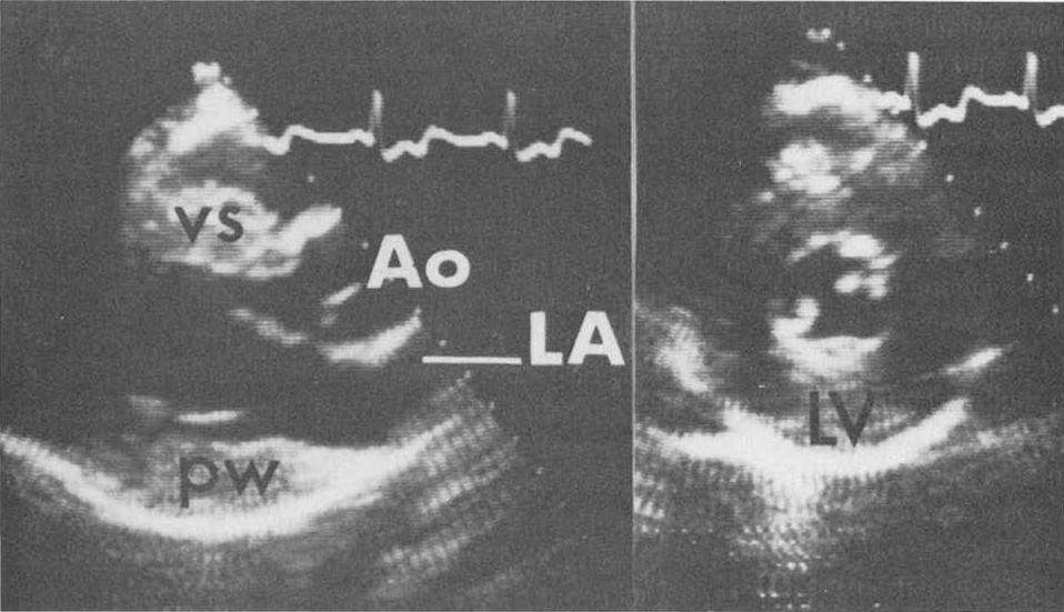 | 958 × 551 (69 KB) | Z3329495 (talk | contribs) | Two-dimensional parasternal long- and short-axis views of an echocardiogram in a 16 year old boy with concentric left ventricular hypertrophy. At age 13 years, his echocardiogram was normal. Ao = aorta; LA = left atrium; LV = left ventricle; pw = posterio |
You cannot overwrite this file.
File usage
The following 2 pages use this file:


