File:Human fetal gonad retinoid receptor expression.jpg
Original file (1,004 × 1,000 pixels, file size: 447 KB, MIME type: image/jpeg)
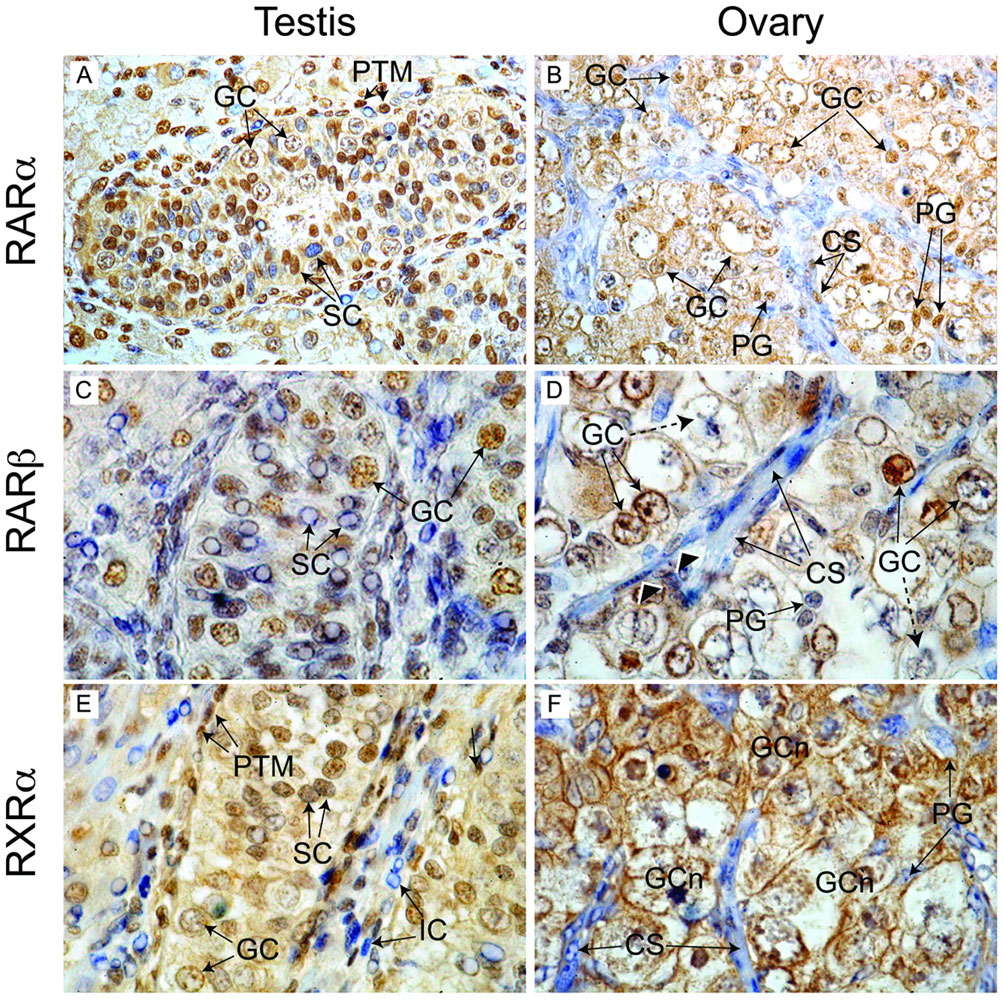
Immunohistochemical localisation of retinoid receptor expression in the human fetal gonad
In the second trimester human fetal testis (A) RARα staining was detected in germ cell (GC) and peritubular myoid (PTM) nuclei. Two populations of Sertoli cells (SC; immunopositive and immunonegative) could also be detected. In the fetal ovary at the same developmental stage (B), RARα expression was detected in the nuclei and cytoplasm of germ cells in nests, and in the nuclei of pregranulosa cells (PG) interspersed between germ cells. Mesenchymal somatic cells in streams (CS) displayed variable staining. RARβ expression was widespread in the fetal testis (C) with all major cell populations displaying intense nuclear staining. In contrast, variable RARβ expression was detected in the germ cells of the fetal ovary (D); with some displaying intensely stained nuclei or both nuclear and cytoplasmic staining (solid arrows) and others showing little or no staining (dashed arrows). Pregranulosa cells were immunonegative, as were somatic cells in streams, although some displayed nuclear staining for RARβ (arrowheads). Peritubular myoid and Sertoli cell nuclei in the testes displayed intense staining for RARβ (E), with weaker expression in detected in germ cells. A population of immunonegative IC was also detected. The distribution of immunostaining for RXRα in the fetal ovary (F) was comparable to that of RARβ, with expression restricted to germ cells in nests (GCn) and absent in somatic cell streams and pregranulosa cells. The widespread nuclear localization of RA receptors in testis suggests cells of all types (including germ cells) are exposed to RA signals. Magnification: 400× (A, B), 1000× (C–F).
Original File Name: Figure 3. Journal.pone.0020249.g003.png (adjust size from original)
Reference
doi:10.1371/journal.pone.0020249.g003
File history
Click on a date/time to view the file as it appeared at that time.
| Date/Time | Thumbnail | Dimensions | User | Comment | |
|---|---|---|---|---|---|
| current | 15:41, 18 June 2011 | | 1,004 × 1,000 (447 KB) | S8600021 (talk | contribs) | ==Immunohistochemical localisation of retinoid receptor expression in the human fetal gonad== In the second trimester human fetal testis (A) RARα staining was detected in germ cell (GC) and peritubular myoid (PTM) nuclei. Two populations of Sertoli cell |
You cannot overwrite this file.
File usage
The following page uses this file: