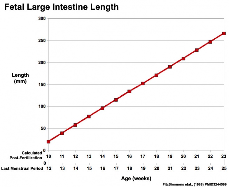
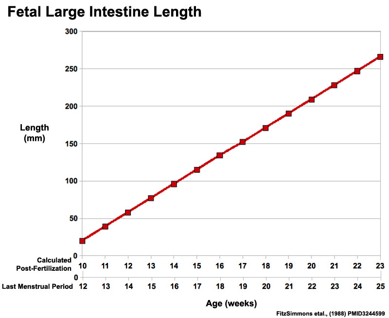
File:Fetal large Intestine length growth graph.jpg
From Embryology
Size of this preview: 735 × 600 pixels.
Original file (800 × 653 pixels, file size: 50 KB, MIME type: image/jpeg)
Human Fetal Small Intestine Length
Graph based on data from FitzSimmons J, Chinn A, Shepard TH. (1988)[1][2]
- 58 fetuses received in the Central Laboratory for Human Embryology between January 1, 1987, and July 1, 1988
- no abnormalities noted on autopsy
- measured from the esophagogastric junction to the pelvic floor
- measurements of stomach, small and large intestines, and appendix length were made and correlated with gestational age as determined by foot length
- overall growth of the gastrointestinal tract and each component was linear with respect to gestational age
- gastrointestinal tracts of aneuploid fetuses fell within the normal range until approximately 20 weeks gestation, after which growth decreased
Note original data based upon gestational age (LMP), post-fertilization age has only been calculated for this current graph.
For educational purposes only.
References
File history
Yi efo/eka'e gwa ebo wo le nyangagi wuncin ye kamina wunga tinya nan
| Gwalagizhi | Nyangagi | Dimensions | User | Comment | |
|---|---|---|---|---|---|
| current | 00:42, 1 Haziran 2011 | | 800 × 653 (50 KB) | S8600021 (talk | contribs) | ==Human Fetal Small Intestine Length== Graph based on data from FitzSimmons J, Chinn A, Shepard TH. (1988)<ref><pubmed>3244599</pubmed></ref><ref><pubmed>16891202</pubmed></ref> * 58 fetuses received in the Central Laboratory for Human Embryology betwee |
You cannot overwrite this file.
File usage
The following 8 pages use this file: