File:Gray1109.jpg
From Embryology
No higher resolution available.
Gray1109.jpg (464 × 487 pixels, file size: 56 KB, MIME type: image/jpeg)
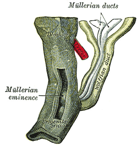
Urogenital Sinus of Female Human Embryo of 8.5 to 9 weeks old
(From model by Keibel)
- Gray's Images: Development | Lymphatic | Neural | Vision | Hearing | Somatosensory | Integumentary | Respiratory | Gastrointestinal | Urogenital | Endocrine | Surface Anatomy | iBook | Historic Disclaimer
| Historic Disclaimer - information about historic embryology pages |
|---|
 |
| iBook - Gray's Embryology | |
|---|---|

|
|
Reference
Gray H. Anatomy of the human body. (1918) Philadelphia: Lea & Febiger.
Cite this page: Hill, M.A. (2024, June 20) Embryology Gray1109.jpg. Retrieved from https://embryology.med.unsw.edu.au/embryology/index.php/File:Gray1109.jpg
- © Dr Mark Hill 2024, UNSW Embryology ISBN: 978 0 7334 2609 4 - UNSW CRICOS Provider Code No. 00098G
File history
Yi efo/eka'e gwa ebo wo le nyangagi wuncin ye kamina wunga tinya nan
| Gwalagizhi | Nyangagi | Dimensions | User | Comment | |
|---|---|---|---|---|---|
| current | 09:27, 28 May 2011 | | 464 × 487 (56 KB) | S8600021 (talk | contribs) | ==Urogenital Sinus of Female Human Embryo of 8.5 to 9 weeks old== (From model by Keibel) {{Gray Anatomy}} Category:Human Category:Genital Category:Female Category:Uterus |
You cannot overwrite this file.
File usage
The following 6 pages use this file: