File:Sabin1909 fig09.jpg
From Embryology
Size of this preview: 495 × 599 pixels. Other resolution: 661 × 800 pixels.
Original file (661 × 800 pixels, file size: 34 KB, MIME type: image/jpeg)
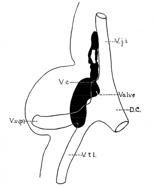
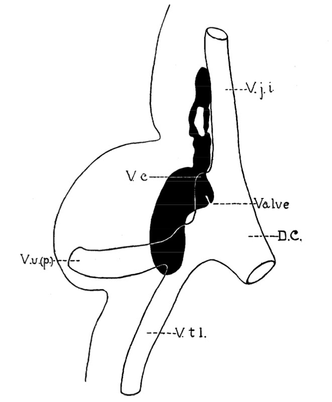
Fig 9. Human embryo measuring 11 mm
Fig. 9. A composite diagram made by superimposing the sections showing the jugular sacs, as shown in Figs. 16 and 17, of human embryo, 11 mm. long, Mall collection. x about 36. D. C., ductus Cuvier ; V., position of valve ; V. j. 1.. vena jugularis interna ; V. t. I., vena thoracicus lateralis ; V. u. (p.) vena ulnarls (primitlva).
Reference
Florence R. Sabin, The lymphatic system in human embryos, with a consideration of the morphology of the system as a whole. American Journal of Anatomy Volume 9, Issue 1, pages 43–91, 1909
File history
Yi efo/eka'e gwa ebo wo le nyangagi wuncin ye kamina wunga tinya nan
| Gwalagizhi | Nyangagi | Dimensions | User | Comment | |
|---|---|---|---|---|---|
| current | 13:54, 30 March 2011 | | 661 × 800 (34 KB) | S8600021 (talk | contribs) | ==Fig 9. Human embryo measuring 11 mm== Fig. 9. A composite diagram made by superimposing the sections showing the jugular sacs, as shown in Figs. 16 and 17, of human embryo, 11 mm. long, Mall collection. x about 36. D. C., ductus Cuvier ; V., position o |
You cannot overwrite this file.
File usage
The following page uses this file: