File:1755-8166-1-21-3-l.jpg
Original file (1,200 × 321 pixels, file size: 28 KB, MIME type: image/jpeg)
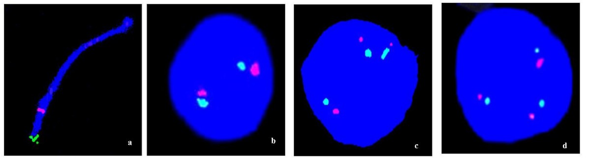
Examples of FISH results on fetal ovarian cells using two chromosome 21-specific probes. a) Location of the probes near the end of the long arm of chromosome 21. b) Normal cell nucleus showing two dual chromosome 21-specific signals. c, d) T21 cell nuclei showing three dual chromosome 21-specific signals.
1755-8166-1-21-3-l.jpg
http://www.molecularcytogenetics.org/content/1/1/21
Hultén et al. Molecular Cytogenetics 2008 1:21 doi:10.1186/1755-8166-1-21
© 2008 Hultén et al; licensee BioMed Central Ltd.
This is an Open Access article distributed under the terms of the Creative Commons Attribution License (http://creativecommons.org/licenses/by/2.0), which permits unrestricted use, distribution, and reproduction in any medium, provided the original work is properly cited.
File history
Yi efo/eka'e gwa ebo wo le nyangagi wuncin ye kamina wunga tinya nan
| Gwalagizhi | Nyangagi | Dimensions | User | Comment | |
|---|---|---|---|---|---|
| current | 22:33, 24 August 2009 | 1,200 × 321 (28 KB) | S8600021 (talk | contribs) | Examples of FISH results on fetal ovarian cells using two chromosome 21-specific probes. a) Location of the probes near the end of the long arm of chromosome 21. b) Normal cell nucleus showing two dual chromosome 21-specific signals. c, d) T21 cell nuclei |
You cannot overwrite this file.
File usage
There are no pages that use this file.