File:Foster037.jpg
Original file (771 × 984 pixels, file size: 123 KB, MIME type: image/jpeg)
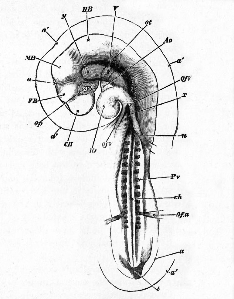
FIG. 37. CHICK OF THE THIRD DAY (FIFTY-FOUR HOURS) VIEWED FROM UNDERNEATH AS A TRANSPARENT OBJECT.
a'. the outer amniotic fold or false amnion. This is very conspicuous around the head, but may also be seen at the tail.
a. the true amnion, very closely enveloping the head, and here seen only between the projections of the several cerebral vesicles. It may also be traced at the tail.
In the embryo of which this is a drawing, the head-fold of the amnion reached a little farther backward than the reference u, but its limit could not be distinctly seen through the body of the embryo. The prominence of the false arnnion at the head is apt to puzzle the student ; but if he bears in mind the fact, which could not well be shewn in Fig. 9, that the whole amniotic fold, both the true and the false limb, is tucked in underneath the head, the matter will on reflection become intelligible.
C. H. cerebral hemisphere. F. B. thalamencephalon or vesicle of the third ventricle. M. B. mid-brain. H. B. hind-brain. Op. optic vesicle. Ot. otic vesicle. Of V. vitelline veins forming the venous roots of the heart. The trunk on the right hand (left trunk when the embryo is viewed in its natural position from above) receives a large branch, shewn by dotted lines, coming from the anterior portion of the sinus terminalis. Ht. the heart, now completely twisted on itself. Ao. the bulbus arteriosus, the three aortic arches being dimly seen stretching from it across the throat, and uniting into the aorta, still more dimly seen as a curved dark line running along the body. The other curved dark line by its side, ending near the reference y, is the notochord ch.
About opposite the line of reference x the aorta divides into two trunks, which, running in the line of the somewhat opaque mesoblastic somites on either side, are not clearly seen. Their branches however, Ofa, the vitelline arteries, are conspicuous and are seen to curve round the commencing side folds.
Pv. mesoblastic somites. Below the level of the vitelline arteries the vertebral plates are but imperfectly cut up into mesoblastic somites, and lower down still, not at all.
x is placed at the "point of divergence" of the splanchnopleure folds. The blind foregut begins here and extends about up to y. x therefore marks the present hind limit of the splanchnopleure folds. The limit of the more transparent somatopleure folds is not shewn.
File history
Yi efo/eka'e gwa ebo wo le nyangagi wuncin ye kamina wunga tinya nan
| Gwalagizhi | Nyangagi | Dimensions | User | Comment | |
|---|---|---|---|---|---|
| current | 09:05, 11 Ocak 2011 | | 771 × 984 (123 KB) | S8600021 (talk | contribs) | FIG. 37. CHICK OF THE THIRD DAY (FIFTY-FOUR HOURS) VIEWED FROM UNDERNEATH AS A TRANSPARENT OBJECT. a'. the outer amniotic fold or false amnion. This is very conspicuous around the head, but may also be seen at the tail. a. the true amnion, very closel |
You cannot overwrite this file.
File usage
The following 2 pages use this file:


