File:Fawcett1905 fig01.jpg
Original file (800 × 1,114 pixels, file size: 145 KB, MIME type: image/jpeg)
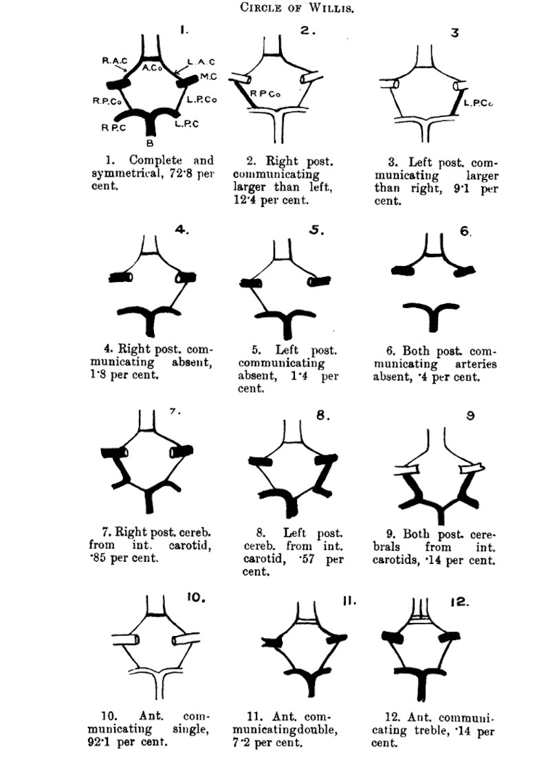
Fig. 1. The circle of Willis
B.—Basilar artery. M.C.—Middle cerebral artery. R. P.C.—Right post. cerebral artery. L, A.C.—Left anterior cerebral, L.P.C.—Lett post. cerebral artery. R. A.C. —Right anterior cerebral. R.P.Co.—Right post, communicating. A.Co.—Anterior communicating. L.P.Co.—Lett post. communicating.
Reference
Fawcett E. and Blachford JV. The circle of Willis - An examination of 700 specimens. (1905) J Anat. Physiol. 40(1): 63.2–70. PMID 17232664
Cite this page: Hill, M.A. (2026, October 4) Embryology Fawcett1905 fig01.jpg. Retrieved from https://embryology.med.unsw.edu.au/embryology/index.php/File:Fawcett1905_fig01.jpg
- © Dr Mark Hill 2026, UNSW Embryology ISBN: 978 0 7334 2609 4 - UNSW CRICOS Provider Code No. 00098G
File history
Yi efo/eka'e gwa ebo wo le nyangagi wuncin ye kamina wunga tinya nan
| Gwalagizhi | Nyangagi | Dimensions | User | Comment | |
|---|---|---|---|---|---|
| current | 12:00, 13 August 2020 | | 800 × 1,114 (145 KB) | Z8600021 (talk | contribs) | |
| 11:58, 13 August 2020 |  | 800 × 1,259 (179 KB) | Z8600021 (talk | contribs) | ===Reference=== {{Ref-Fawcett1905}} |
You cannot overwrite this file.
File usage
The following page uses this file:


