File:HamiltonBoyd1960 plate11.jpg
Original file (1,280 × 1,707 pixels, file size: 659 KB, MIME type: image/jpeg)
Plate 11
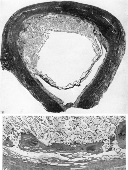
Fig. 29. Photograph ( x 1-8) of a uterus which contained a 60 mm. foetus (CX. 112). The uterine lumen is still apparent in the lower half of the uterus. The decidua basalis is very thin. Distorted and dilated uterine glands are present in the decidua vera to the right of the area of placental attachment.
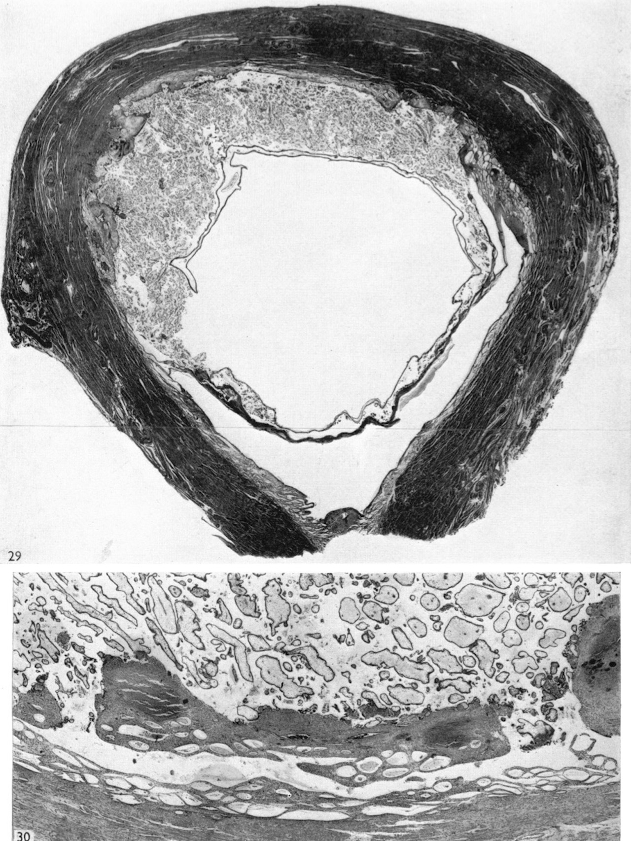
Fig. 30. Photomicrograph ( x 18) of a section through the central portion of the in situ placenta of a 30 mm. embryo (CX. 106). Two large openings from the intervillous space into uterine veins are shown. A plug of villi can be seen in the right of these communications.
File history
Yi efo/eka'e gwa ebo wo le nyangagi wuncin ye kamina wunga tinya nan
| Gwalagizhi | Nyangagi | Dimensions | User | Comment | |
|---|---|---|---|---|---|
| current | 13:51, 6 August 2020 | | 1,280 × 1,707 (659 KB) | Z8600021 (talk | contribs) | |
| 13:50, 6 August 2020 |  | 1,030 × 1,429 (465 KB) | Z8600021 (talk | contribs) |
You cannot overwrite this file.
File usage
The following 12 pages use this file:
- Paper - Development of the human placenta in the first three months of gestation (1960)
- File:HamiltonBoyd1960 fig02.jpg
- File:HamiltonBoyd1960 fig03.jpg
- File:HamiltonBoyd1960 fig04.jpg
- File:HamiltonBoyd1960 fig05.jpg
- File:HamiltonBoyd1960 fig06.jpg
- File:HamiltonBoyd1960 fig07.jpg
- File:HamiltonBoyd1960 fig08.jpg
- File:HamiltonBoyd1960 plate02.jpg
- File:HamiltonBoyd1960 plate03.jpg
- File:HamiltonBoyd1960 plate13.jpg
- Template:HamiltonBoyd1960 plates