File:Marshall1881 plate01.jpg
Original file (941 × 1,493 pixels, file size: 455 KB, MIME type: image/jpeg)
Summary
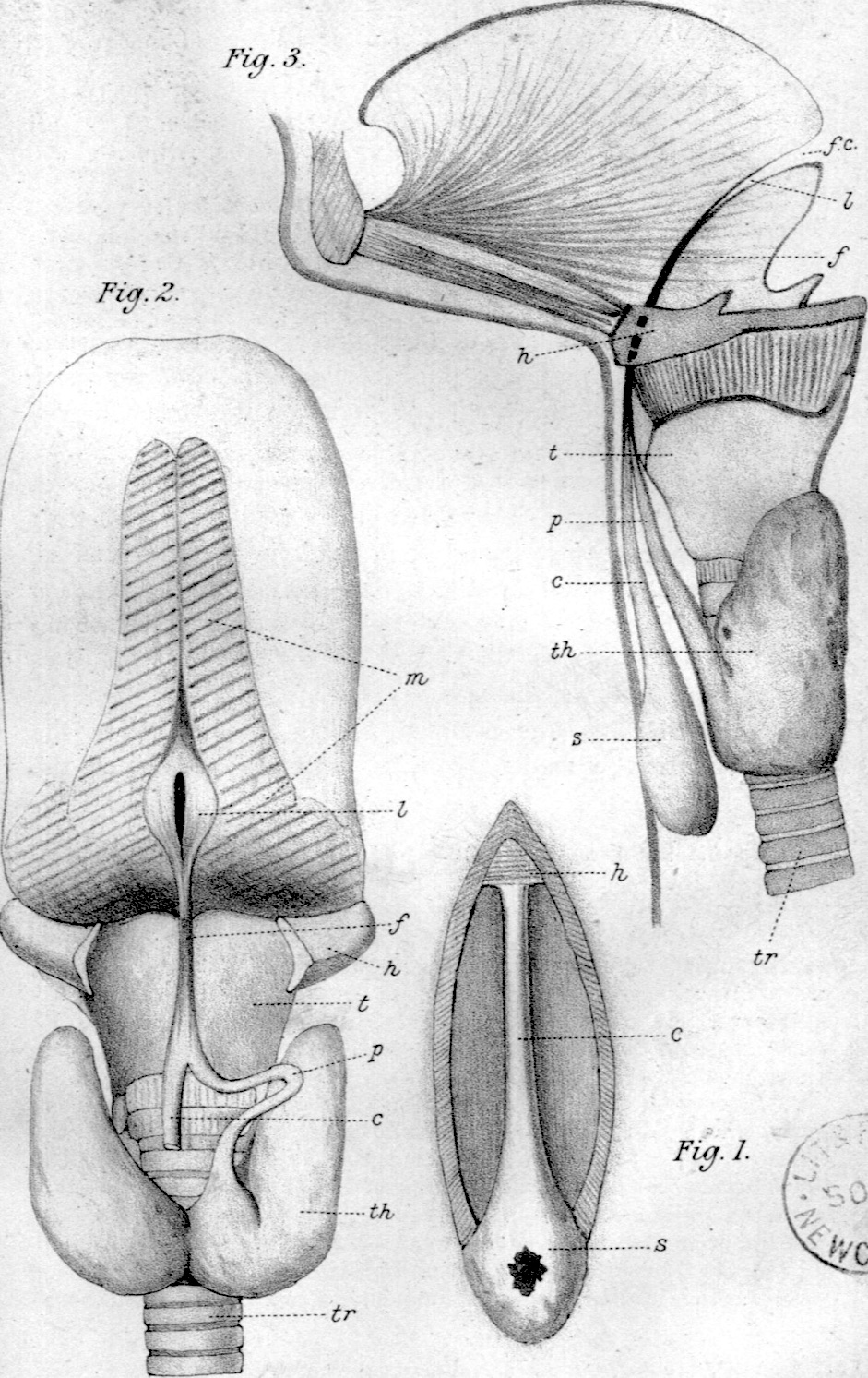
Explanation of Plate I
(All the figures are natural size.)
Fig. 1. Canal of His exposed by dissecting the skin from the median line of the neck. c¢, canal; 2, hyoid bone; s, sac with open sinus.
Fig. 2. Dissection of the thyro-glossal duct, thyroid gland, and base of the tongue. , upper end of canal of His; f, fibrous cord; A, hyoid bone; J, lingual duct opened and fixed by pins; m, muscles cut through at base of tongue; p, pyramidal lobe; z, thyroid cartilage; ¢h, thyroid gland ; tr, trachea. ;
Fig. 3. Semi-diagrammatic figure: the upper parts are in vertical section, the lower part in elevation; /f. c, foramen cecum; s, sac with sinus opening in front of neck ; other letters as before,
1 Loc, cit., fig. 67.
Reference
Marshall AM. Thyro-Glossal duct or “canal of His”. (1881) J Anat. Physiol. 16(3): 305–354. PMID 17231961
Cite this page: Hill, M.A. (2026, October 1) Embryology Marshall1881 plate01.jpg. Retrieved from https://embryology.med.unsw.edu.au/embryology/index.php/File:Marshall1881_plate01.jpg
- © Dr Mark Hill 2026, UNSW Embryology ISBN: 978 0 7334 2609 4 - UNSW CRICOS Provider Code No. 00098G
File history
Yi efo/eka'e gwa ebo wo le nyangagi wuncin ye kamina wunga tinya nan
| Gwalagizhi | Nyangagi | Dimensions | User | Comment | |
|---|---|---|---|---|---|
| current | 09:27, 22 July 2020 | | 941 × 1,493 (455 KB) | Z8600021 (talk | contribs) | |
| 09:24, 22 July 2020 | 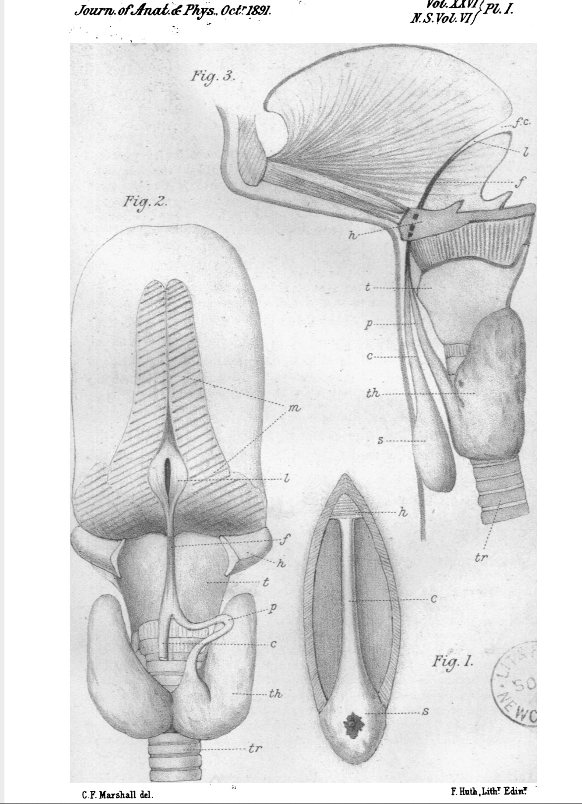 | 1,177 × 1,626 (415 KB) | Z8600021 (talk | contribs) | ==Explanation of Plate I== (All the figures are natural size.) Fig. 1. Canal of His exposed by dissecting the skin from the median line of the neck. c¢, canal; 2, hyoid bone; s, sac with open sinus. Fig. 2. Dissection of the thyro-glossal duct, thyroid gland, and base of the tongue. , upper end of canal of His; f, fibrous cord; A, hyoid bone; J, lingual duct opened and fixed by pins; m, muscles cut through at base of tongue; p, pyramidal lobe; z, thyroid cartilage; ¢h, thyroid gland ; tr,... |
You cannot overwrite this file.
File usage
The following page uses this file:


