File:Human-retina-01.jpg
From Embryology
Size of this preview: 800 × 492 pixels. Other resolution: 1,000 × 615 pixels.
Original file (1,000 × 615 pixels, file size: 197 KB, MIME type: image/jpeg)
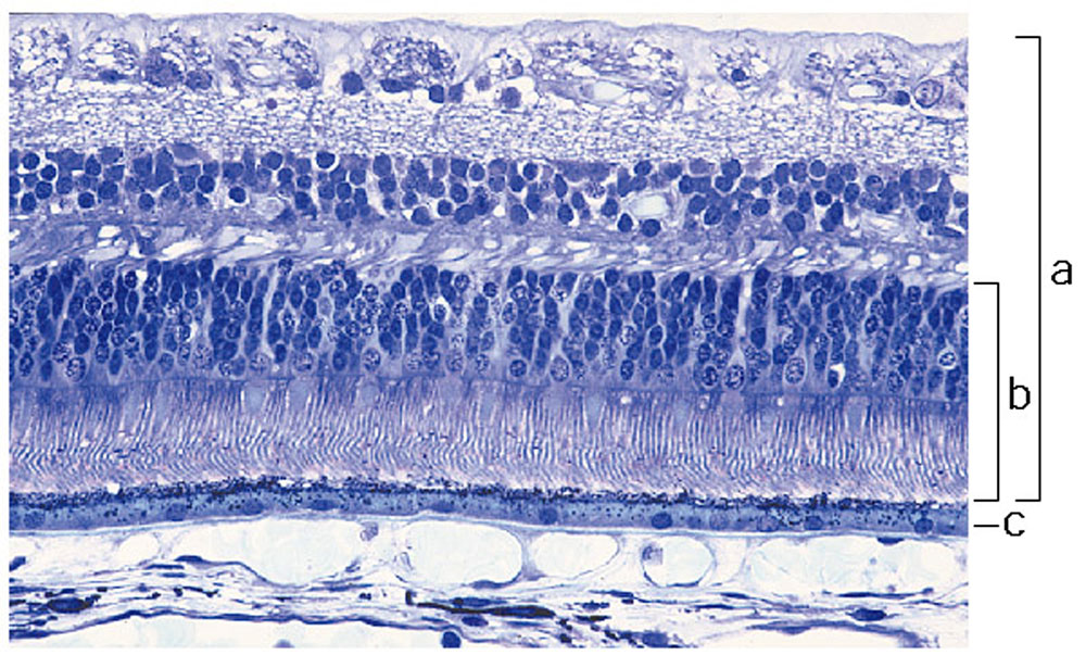
Adult Human Retina Histology
Light micrograph of normal human retina stained with Richardson's methylene blue/azure II. Light enters the retina from the top of the image.
(a) Neural retina
(b) photoreceptor layer
(c) retinal pigment epithelium (RPE)
From the Human Retina Teaching Set, Scheie Eye Institute, University of Pennsylvania, Philadelphia, USA. Courtesy of Ann Milam.
Original Image Name: Figure 1. http://genomebiology.com/2002/3/8/reviews/1022/figure/F1
Reference
<pubmed>12186651</pubmed>
File history
Yi efo/eka'e gwa ebo wo le nyangagi wuncin ye kamina wunga tinya nan
| Gwalagizhi | Nyangagi | Dimensions | User | Comment | |
|---|---|---|---|---|---|
| current | 14:56, 16 October 2010 | | 1,000 × 615 (197 KB) | S8600021 (talk | contribs) | ==Adult Human Retina Histology== Light micrograph of normal human retina stained with Richardson's methylene blue/azure II. Light enters the retina from the top of the image. (a) Neural retina (b) photoreceptor layer (c) retinal pigment epithelium (RP |
You cannot overwrite this file.
File usage
The following 2 pages use this file: