File:Lockwood1887b fig29.jpg

Original file (800 × 656 pixels, file size: 121 KB, MIME type: image/jpeg)
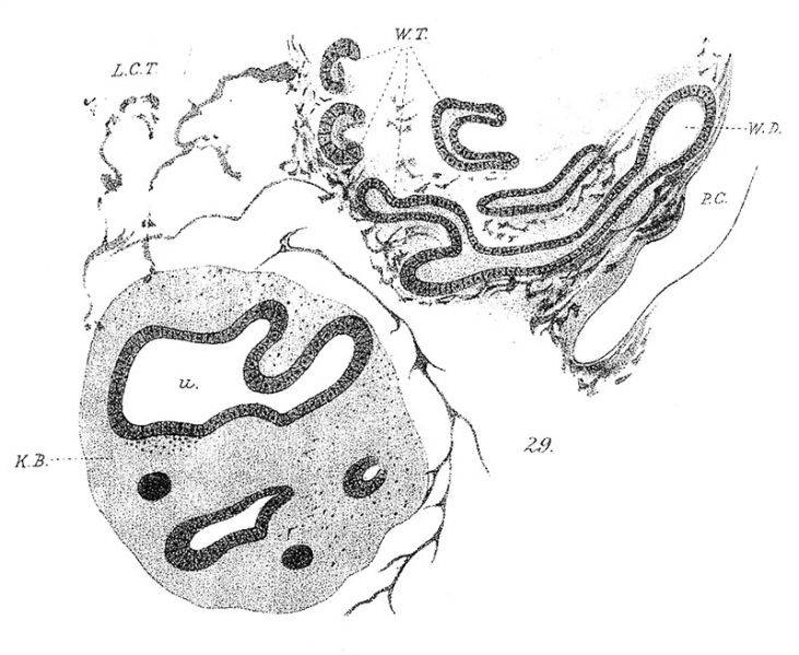
Plate II
Fig. 29. Kidney and hinder part of the Wolffian body of a rabbit of thirteenth day. AB, kidney blastema; U, ureter; LCT, loose tissue, which surrounds kidney blastema ; WD, Wolffian duct; W7;, Wolffian tubules; PC, peritoneal cavity. x 70.
Reference
Lockwood CB. Development and transition of the testis, normal and abnormal. (1887) J Anat. 22(1): 38-77. PMID 17231729
Cite this page: Hill, M.A. (2024, June 26) Embryology Lockwood1887b fig29.jpg. Retrieved from https://embryology.med.unsw.edu.au/embryology/index.php/File:Lockwood1887b_fig29.jpg
- © Dr Mark Hill 2024, UNSW Embryology ISBN: 978 0 7334 2609 4 - UNSW CRICOS Provider Code No. 00098G
File history
Yi efo/eka'e gwa ebo wo le nyangagi wuncin ye kamina wunga tinya nan
| Gwalagizhi | Nyangagi | Dimensions | User | Comment | |
|---|---|---|---|---|---|
| current | 19:05, 14 April 2020 | | 800 × 656 (121 KB) | Z8600021 (talk | contribs) |
You cannot overwrite this file.
File usage
The following page uses this file: