File:Lockwood1888a fig47.jpg
From Embryology
Size of this preview: 800 × 570 pixels. Other resolution: 1,000 × 712 pixels.
Original file (1,000 × 712 pixels, file size: 279 KB, MIME type: image/jpeg)
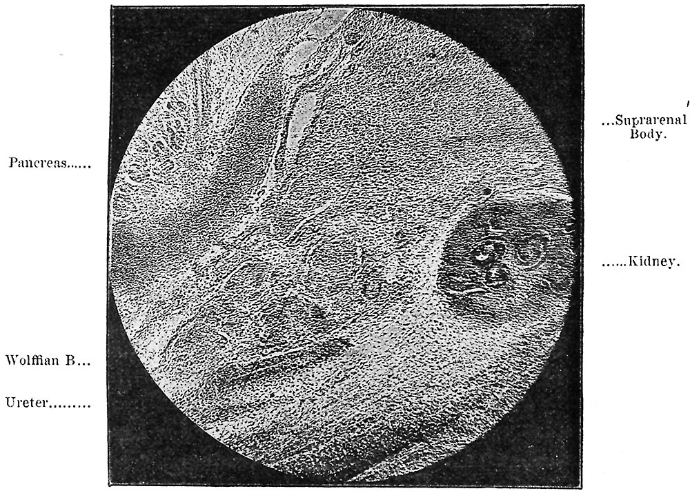
Fig. 47. Continuity of Wolffian and Suprarenal Bodies in a Human Embryo of the seventh week
Electrotype from photograph of section.
Reference
Lockwood CB. Development and transition of the testis, normal and abnormal. (1888) J Anat. 22(3): 460.1-478. PMID 7231755
Cite this page: Hill, M.A. (2026, October 4) Embryology Lockwood1888a fig47.jpg. Retrieved from https://embryology.med.unsw.edu.au/embryology/index.php/File:Lockwood1888a_fig47.jpg
- © Dr Mark Hill 2026, UNSW Embryology ISBN: 978 0 7334 2609 4 - UNSW CRICOS Provider Code No. 00098G
File history
Yi efo/eka'e gwa ebo wo le nyangagi wuncin ye kamina wunga tinya nan
| Gwalagizhi | Nyangagi | Dimensions | User | Comment | |
|---|---|---|---|---|---|
| current | 14:28, 14 April 2020 | | 1,000 × 712 (279 KB) | Z8600021 (talk | contribs) | resize |
| 14:23, 14 April 2020 | 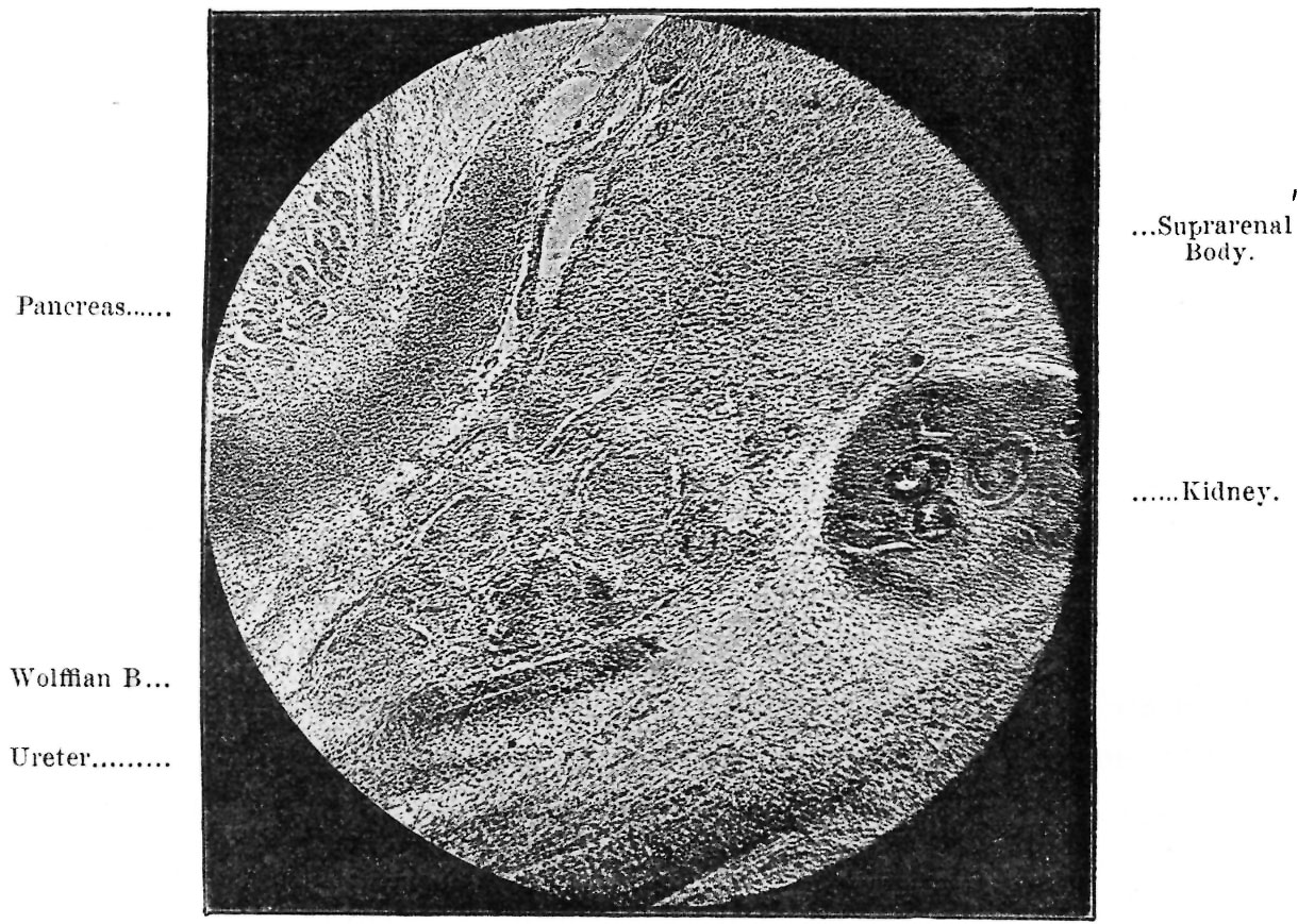 | 1,220 × 869 (353 KB) | Z8600021 (talk | contribs) | crop | |
| 14:18, 14 April 2020 |  | 1,279 × 1,017 (322 KB) | Z8600021 (talk | contribs) | Lockwood1888a fig47.jpg |
You cannot overwrite this file.
File usage
The following page uses this file:


