File:Johnston1907 fig018.jpg
From Embryology
Size of this preview: 601 × 600 pixels. Other resolution: 1,000 × 998 pixels.
Original file (1,000 × 998 pixels, file size: 119 KB, MIME type: image/jpeg)
Summary
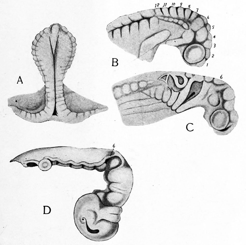
Fig. 18. Four stages in the development of the selachian brain to show the history of the neuromeres. After Locy. In A, B and C the whole head is shown, in D the brain alone. The Arabic numerals indicate the neuromeres.
File history
Yi efo/eka'e gwa ebo wo le nyangagi wuncin ye kamina wunga tinya nan
| Gwalagizhi | Nyangagi | Dimensions | User | Comment | |
|---|---|---|---|---|---|
| current | 23:40, 23 February 2020 | | 1,000 × 998 (119 KB) | Z8600021 (talk | contribs) | |
| 23:34, 23 February 2020 | 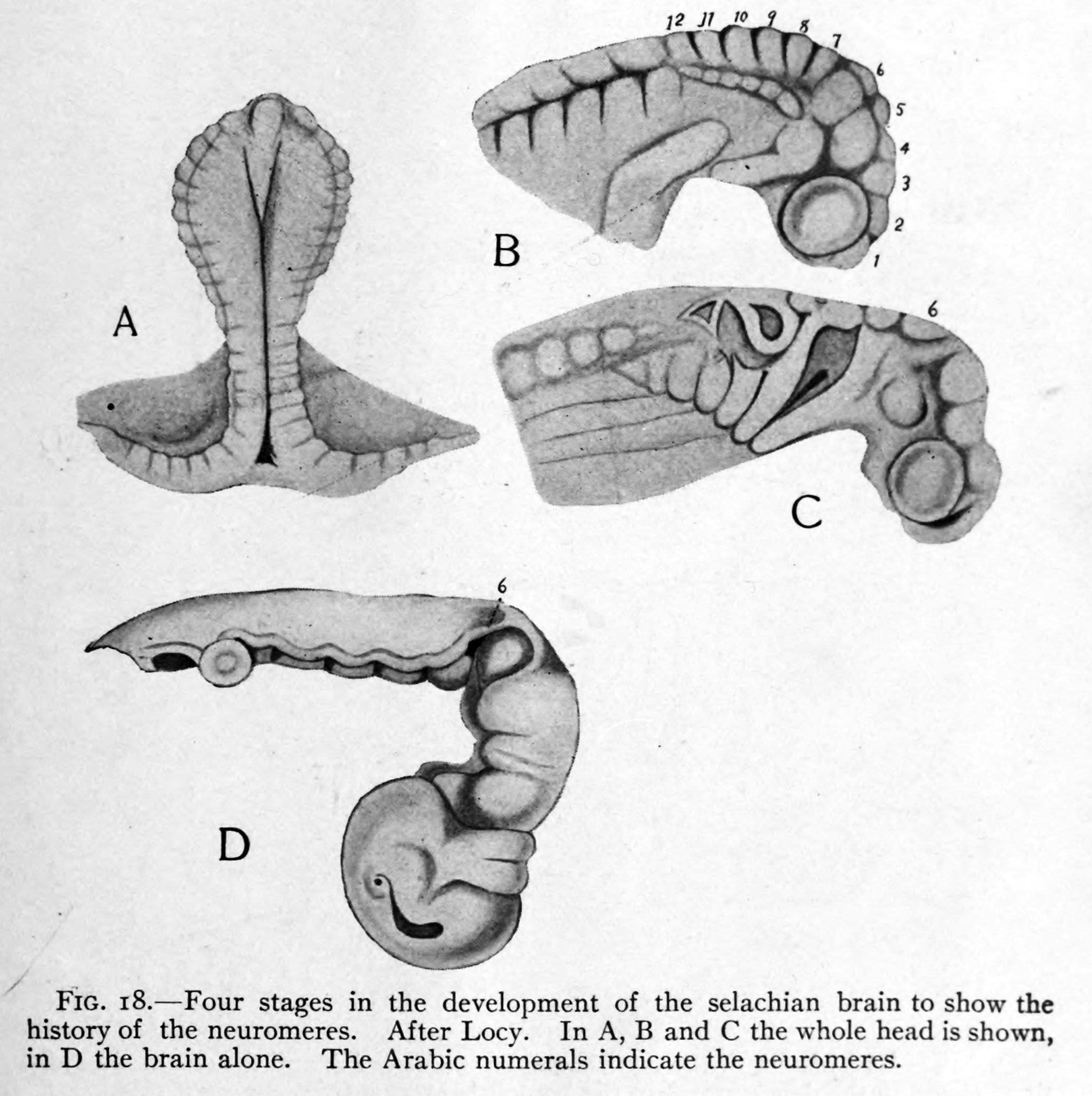 | 2,106 × 2,113 (429 KB) | Z8600021 (talk | contribs) | Fig. 18. Four stages in the development of the selachian brain to show the history of the neuromeres. After Locy. In A, B and C the whole head is shown, in D the brain alone. The Arabic numerals indicate the neuromeres. |
You cannot overwrite this file.
File usage
The following 2 pages use this file:


