File:Flecker1932 fig07.jpg

Flecker1932_fig07.jpg (700 × 506 pixels, file size: 58 KB, MIME type: image/jpeg)
Fig. 7. Male, age 18. Epiphyses along perineal margin at apex of perineal triangle
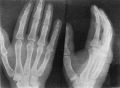
Fig. 6. Age 21. Sesamoids at heads of first, second, third and fifth metacarpals.
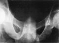
Fig. 7. Male, age 18. Epiphyses along perineal margin at apex of perineal triangle.
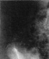
Fig. 8. Female, age 14. Epiphysis for anterior superior iliac spine.
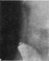
Fig. 9. Female, age 17. Epiphysis for anterior superior iliac spine.
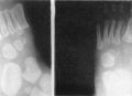
Fig. 10. Female, aged 2 years and 3 months. Two centres present for first cuneiform.


Reference
Flecker H. (1932). Roentgenographic observations of the times of appearance of epiphyses and their fusion with the diaphyses. (1932) J Anat. 67: 118-164.3 PMID 17104405
Cite this page: Hill, M.A. (2026, October 1) Embryology Flecker1932 fig07.jpg. Retrieved from https://embryology.med.unsw.edu.au/embryology/index.php/File:Flecker1932_fig07.jpg
- © Dr Mark Hill 2026, UNSW Embryology ISBN: 978 0 7334 2609 4 - UNSW CRICOS Provider Code No. 00098G
File history
Yi efo/eka'e gwa ebo wo le nyangagi wuncin ye kamina wunga tinya nan
| Gwalagizhi | Nyangagi | Dimensions | User | Comment | |
|---|---|---|---|---|---|
| current | 10:49, 9 February 2020 | | 700 × 506 (58 KB) | Z8600021 (talk | contribs) |
You cannot overwrite this file.
File usage
The following 7 pages use this file:


