File:Fetal integumentary histology 01.jpg
From Embryology
No higher resolution available.
Fetal_integumentary_histology_01.jpg (800 × 219 pixels, file size: 74 KB, MIME type: image/jpeg)
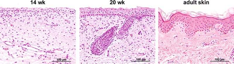
Histology of 14 and 20 weeks’ gestation fetal skin and of adult skin
Fetal integumentary histology
- At 14 weeks, the epidermis consisted of a basal layer, an intermediate cell layer and periderm.
- At 20 weeks, the number of intermediate cells layers was increased and developing hair follicles were visible.
- The adult skin contained basal, spinous, granular and cornified layers.
Scale bars 100 μm
Original file name: Fig. 1 403_2009_989_Fig1_HTML.gif (original figure increased in size)
Reference
<pubmed>19701759</pubmed>| PMC2799629 | [ http://www.springerlink.com/content/lv415257322x8247/fulltext.html Arch Dermatol Res]
© Coolen NA, Schouten KC, Middelkoop E, Ulrich MM. 2009 Open Access - This article is distributed under the terms of the Creative Commons Attribution Noncommercial License which permits any noncommercial use, distribution, and reproduction in any medium, provided the original author(s) and source are credited.
File history
Yi efo/eka'e gwa ebo wo le nyangagi wuncin ye kamina wunga tinya nan
| Gwalagizhi | Nyangagi | Dimensions | User | Comment | |
|---|---|---|---|---|---|
| current | 00:44, 10 October 2010 | 800 × 219 (74 KB) | S8600021 (talk | contribs) | ==Histology of 14 and 20 weeks’ gestation fetal skin and of adult skin== ==Fetal integumentary histology== * At 14 weeks, the epidermis consisted of a basal layer, an intermediate cell layer and periderm. * At 20 weeks, the number of intermediate cell |
You cannot overwrite this file.