File:MERS-CoV EM1.jpg
From Embryology
No higher resolution available.
MERS-CoV_EM1.jpg (537 × 537 pixels, file size: 68 KB, MIME type: image/jpeg)
Summary
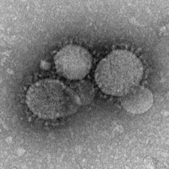
MERS-CoV particles as seen by negative stain electron microscopy. Virions contain characteristic club-like projections emanating from the viral membrane.
Reference
CDC Source: Cynthia Goldsmith/Maureen Metcalfe/Azaibi Tamin
Cite this page: Hill, M.A. (2026, September 30) Embryology MERS-CoV EM1.jpg. Retrieved from https://embryology.med.unsw.edu.au/embryology/index.php/File:MERS-CoV_EM1.jpg
- © Dr Mark Hill 2026, UNSW Embryology ISBN: 978 0 7334 2609 4 - UNSW CRICOS Provider Code No. 00098G
File history
Yi efo/eka'e gwa ebo wo le nyangagi wuncin ye kamina wunga tinya nan
| Gwalagizhi | Nyangagi | Dimensions | User | Comment | |
|---|---|---|---|---|---|
| current | 00:31, 21 Ocak 2020 | | 537 × 537 (68 KB) | Z8600021 (talk | contribs) | MERS-CoV particles as seen by negative stain electron microscopy. Virions contain characteristic club-like projections emanating from the viral membrane. ===Reference=== CDC Source: Cynthia Goldsmith/Maureen Metcalfe/Azaibi Tamin {{Footer}} Category:VirusCategory:EM |
You cannot overwrite this file.
File usage
The following 3 pages use this file:


