Talk:2010 Lab 10
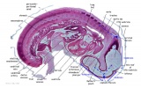
Stage 13

|

|

|

|
| B1L Dorsal portion of hypopharyngeal eminence. Rathke's pouch derived from ectoderm anterior to the buccopharyngeal membrane ( rudimentary adenohypophysis). | B2L Dorsal portion of hypopharyngeal eminence. Rathke's pouch derived from ectoderm anterior to the buccopharyngeal membrane ( rudimentary adenohypophysis). | B3L Rudimentary thyroid ventral to aortic sac (also seen in B2, ventral to the hypopharyngeal eminence). | G7L (Close to midline in head region). Forebrain, midbrain, hindbrain (with thin roof). Arrowed Rathke's pouch. Floor of pharynx with foramen caecum (the tongue has not yet formed). |
Stage 22

|
A6L Thalamus/hypothalamus. |

|
B2L Neurohyophysis and adenohypophysis. Remnant of Rathke's pouch (residual lumen) in adenohypophysis |

|
B3L Adenohypophysis. |

|
C3L Glottic region with cricoid cartilage and descending process of thyroid cartilage laterally. |

|
C4L Section damaged, but shows thyroid gland lateral to trachea. |

|
C5L Thyroid gland. |

|
D1L Thymus gland. |

|
E6L R,L adrenal glands under diaphragm |

|
E7L Large adrenal glands. |

|
F1L Adrenal glands. R. Kidney. Autonomic ganglia (partly the adrenal medulla precursors). |

|
F2L Kidneys (note retroperitoneal location). Cortex. Medulla. L. Adrenal gland. |
List of serial section images containing endocrine components.
A6: Thalamus/hypothalamus.
B2: Neurohyophysis and adenohypophysis. Remnant of Rathke's pouch (residual lumen) in adenohypophysis.
B3: Adenohypophysis.
C3: Glottic region with cricoid cartilage and descending process of thyroid cartilage laterally.
C4: Section damaged, but shows thyroid gland lateral to trachea.
C5: Thyroidgland.
Dl: Thymus gland.
E6: R,L adrenal glands under diaphragm.
E7: Large adrenal glands.
Fl: Adrenal glands. R. Kidney. Autonomic ganglia (partly the adrenal medulla precursors).
F2: Kidneys (note retroperitoneal location). Cortex. Medulla. L. Adrenal gland.
Stage 22 Embryo (Human) Selected High power
Hypophysis (pituitary)
A7: Overview of embryo (stage 22) head region where high power sections is from in B2. Hypophysis (pituitary) in centre of image cut tranversely.
B2: Neurohypophysis (infundibular part with hypothalamic recess of 3rd ventricle). Cavity in centre of image is the residual lumen of Rathke's pouch, which will form pars intermedia. Surrounding the hypophysis is sphenoid cartilage.
Thyroid
C2: Overview of embryo (stage 22) neck region where high power sections is from in C3. Thyroid gland at top of image lying anterior and extending lateral to the trachea.
C3: Thyroid gland.
Thymus
C5: Overview of embryo (stage 22) chest region where high power sections is from in C6. Thymus gland at top of image anterior to section through aortic arch.
C6: Thymus gland.
Adrenal
E6: Overview of embryo (stage 22) at the level of the liver where adrenals are seen.
F3: Fetal and permanent adrenal cortex. The medulla of the adrenal gland is of neural crest origin and it is not yet encapsulated by the cortex.