File:Thyroid-development-cartoon.jpg
From Embryology
No higher resolution available.
Thyroid-development-cartoon.jpg (600 × 541 pixels, file size: 32 KB, MIME type: image/jpeg)
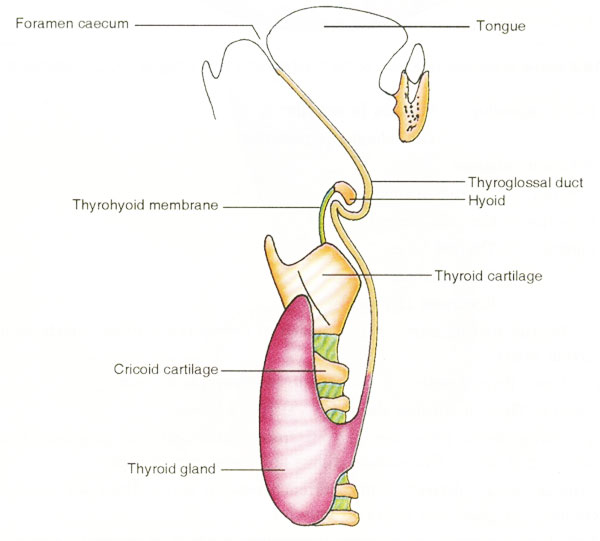
Cartoon showing the development of thyroid gland
- The thyroid gland begins to develop as a median thickening of endoderm on the floor of the pharynx between the first and second pharyngeal pouches.
- This area later invaginates to form the median diverticulum, which appears in the later half of the fourth week.
- This thyroid diverticulum grows further, becoming a solid cellular cord called the thyroglossal duct.
- The duct grows caudally and bifurcates to give rise to the thyroid lobes and the isthmus.
Original file name: Figure 4 http://www.ncbi.nlm.nih.gov/pmc/articles/PMC2827060/figure/F4/
Reference
<pubmed>20181171</pubmed>| PMC2827060
This is an Open Access article distributed under the terms of the Creative Commons Attribution License (http://creativecommons.org/licenses/by/3.0), which permits unrestricted use, distribution, and reproduction in any medium, provided the original work is properly cited.
File history
Yi efo/eka'e gwa ebo wo le nyangagi wuncin ye kamina wunga tinya nan
| Gwalagizhi | Nyangagi | Dimensions | User | Comment | |
|---|---|---|---|---|---|
| current | 23:20, 5 October 2010 | | 600 × 541 (32 KB) | S8600021 (talk | contribs) | Showing the development of thyroid gland. The thyroid gland begins to develop as a median thickening of endoderm on the floor of the pharynx between the first and second pharyngeal pouches. This area later invaginates to form the median diverticulum, whi |
You cannot overwrite this file.
File usage
The following 7 pages use this file: