File:Bradley1908 fig01.jpg

Original file (1,000 × 836 pixels, file size: 87 KB, MIME type: image/jpeg)
Fig. 1. Outline of model of liver of 6 mm pig embryo (Caudal surface)
d., left laterai lobe; 7.U.., right lateral lobe: c.l., central lobe; g.f., fossa for stomach; J.u.v., left umbilical vein; 7.u.v., right umbilical vein; r.o-m.v., right omphalo-mesenteric vein ; g.b., gall-bladder. The dotted area is not covered by peritoneum.
Reference
Bradley OC. A contribution to the morphology and development of the mammalian liver. (1908) J Anat. 43: 1-42. PMID 17232788
Cite this page: Hill, M.A. (2024, June 3) Embryology Bradley1908 fig01.jpg. Retrieved from https://embryology.med.unsw.edu.au/embryology/index.php/File:Bradley1908_fig01.jpg
- © Dr Mark Hill 2024, UNSW Embryology ISBN: 978 0 7334 2609 4 - UNSW CRICOS Provider Code No. 00098G
File history
Click on a date/time to view the file as it appeared at that time.
| Date/Time | Thumbnail | Dimensions | User | Comment | |
|---|---|---|---|---|---|
| current | 20:39, 21 November 2019 | | 1,000 × 836 (87 KB) | Z8600021 (talk | contribs) | |
| 20:36, 21 November 2019 | 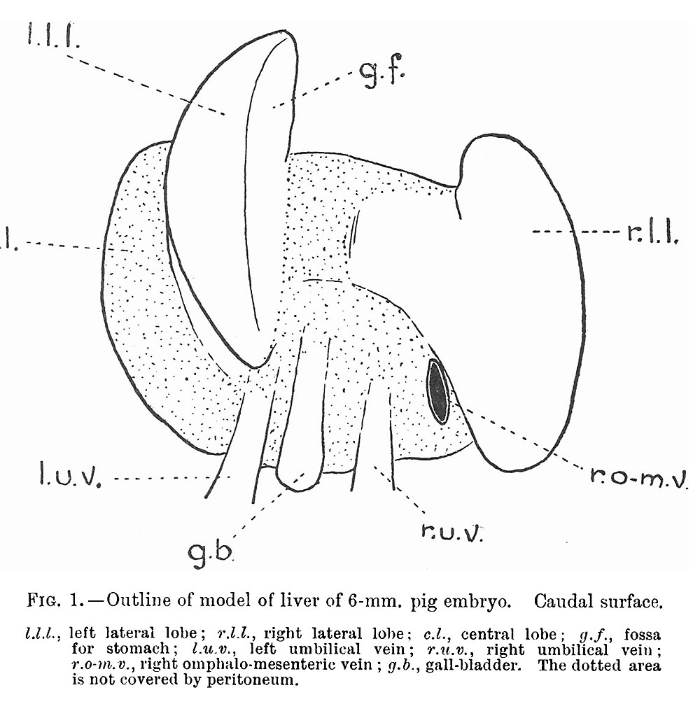 | 1,000 × 1,006 (132 KB) | Z8600021 (talk | contribs) | ===Reference=== {{Ref-Bradley1908}} |
You cannot overwrite this file.
File usage
The following 2 pages use this file: