File:Murine Cereblon in complex with Thalidomide.jpg
From Embryology
Size of this preview: 743 × 599 pixels. Other resolution: 1,086 × 876 pixels.
Original file (1,086 × 876 pixels, file size: 120 KB, MIME type: image/jpeg)
Summary
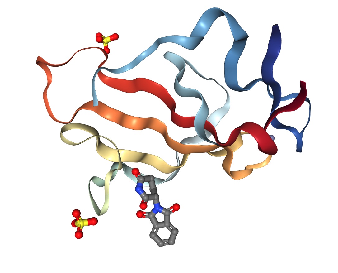
Crystal Structure of Murine Cereblon in Complex with Thalidomide
Reference
Cite images created with the PDB ID and associated publication, NGL Viewer (AS Rose et al. (2018) NGL viewer: web-based molecular graphics for large complexes. Bioinformatics doi:10.1093/bioinformatics/bty419), and RCSB PDB.
https://www.rcsb.org/3d-view/4TZC/1
Cite this page: Hill, M.A. (2026, October 5) Embryology Murine Cereblon in complex with Thalidomide.jpg. Retrieved from https://embryology.med.unsw.edu.au/embryology/index.php/File:Murine_Cereblon_in_complex_with_Thalidomide.jpg
- © Dr Mark Hill 2026, UNSW Embryology ISBN: 978 0 7334 2609 4 - UNSW CRICOS Provider Code No. 00098G
File history
Yi efo/eka'e gwa ebo wo le nyangagi wuncin ye kamina wunga tinya nan
| Gwalagizhi | Nyangagi | Dimensions | User | Comment | |
|---|---|---|---|---|---|
| current | 14:16, 11 April 2019 | | 1,086 × 876 (120 KB) | Z8600021 (talk | contribs) | ==Crystal Structure of Murine Cereblon in Complex with Thalidomide== ===Reference=== Cite images created with the PDB ID and associated publication, NGL Viewer (AS Rose et al. (2018) NGL viewer: web-based molecular graphics for large complexes. Bioinf... |
You cannot overwrite this file.
File usage
The following page uses this file:


