File:Aorta coarctation echocardiogram.jpg
From Embryology
No higher resolution available.
Aorta_coarctation_echocardiogram.jpg (601 × 283 pixels, file size: 28 KB, MIME type: image/jpeg)
Summary
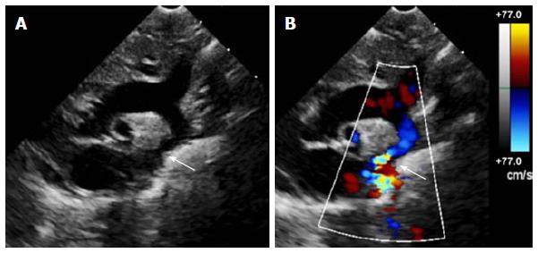
Echocardiogram of coarctation
A: Two-dimensional transthoracic echocardiogram image obtained from the suprasternal notch in an 11-day-old infant demonstrating discrete coarctation (arrow);
B: Colour Doppler of the same image with aliasing of flow at the site of coarctation (arrow).
Figure 1 WJC-7-765-g001.jpg
File history
Click on a date/time to view the file as it appeared at that time.
| Date/Time | Thumbnail | Dimensions | User | Comment | |
|---|---|---|---|---|---|
| current | 14:40, 8 March 2019 | | 601 × 283 (28 KB) | Z8600021 (talk | contribs) | ==Echocardiogram of coarctation== A: Two-dimensional transthoracic echocardiogram image obtained from the suprasternal notch in an 11-day-old infant demonstrating discrete coarctation (arrow); B: Colour Doppler of the same image with aliasing of flo... |
You cannot overwrite this file.
File usage
The following page uses this file: