File:B lymphocyte EM08.jpg
Original file (1,000 × 730 pixels, file size: 111 KB, MIME type: image/jpeg)
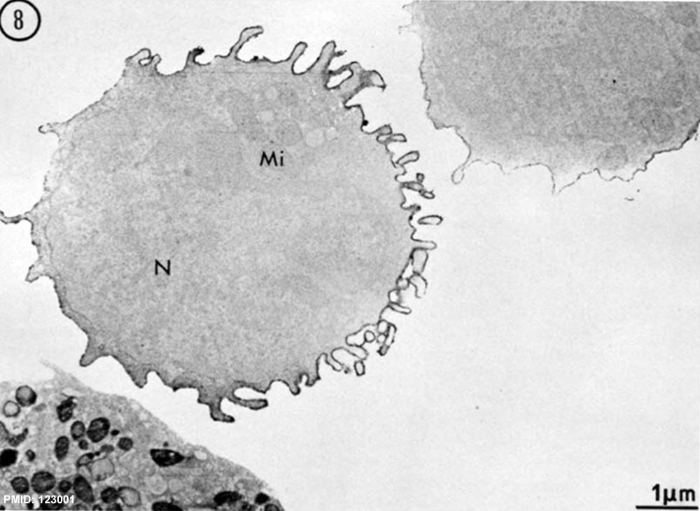
Human B Lymphocyte Electron Micrograph
Fig. 8 Normal B lymphocyte with surface IgM. Microvilli are flowing toward one pole of the cell, the remaining membrane being relatively smoother and moderately labeled. An intermediate B lymphocyte is seen at upper right, and a neutrophil polymorphonuclear at lower left. The latter exhibits peroxidase-containing granules; its membrane is not labeled.x 12,000
- N, nucleus
- Mi, mitochondria
- Lymphocyte EM Images: T and B Lymphocytes 1 TEM | T and B Lymphocytes 2 TEM | T Lymphocyte SEM | B lymphocyte 1 TEM | B lymphocyte 2 TEM | B lymphocyte 3 TEM | Plasma Cell TEM | T2 Lymphocyte 1 TEM | T2 Lymphocyte 2 TEM | lymphocyte rosettes | T lymphocyte 1 | T lymphocyte 2 | T lymphocyte 3 | T lymphocyte 4 | T lymphocyte 5 | T lymphocyte 6 | B lymphocyte | B lymphocytes TEM | Immune System Development | Blood
References
Lejonc RJ, Gourdin MF, Mannoni P, Dreyfus B & Reyes F. (1975). The surface morphology of human B lymphocytes as revealed by immunoelectron microscopy. J. Exp. Med. , 141, 392-410. PMID: 123001
Copyright
Rockefeller University Press - Copyright Policy This article is distributed under the terms of an Attribution–Noncommercial–Share Alike–No Mirror Sites license for the first six months after the publication date (see http://www.jcb.org/misc/terms.shtml). After six months it is available under a Creative Commons License (Attribution–Noncommercial–Share Alike 4.0 Unported license, as described at https://creativecommons.org/licenses/by-nc-sa/4.0/ ). (More? Help:Copyright Tutorial)
Cite this page: Hill, M.A. (2024, June 15) Embryology B lymphocyte EM08.jpg. Retrieved from https://embryology.med.unsw.edu.au/embryology/index.php/File:B_lymphocyte_EM08.jpg
- © Dr Mark Hill 2024, UNSW Embryology ISBN: 978 0 7334 2609 4 - UNSW CRICOS Provider Code No. 00098G
File history
Click on a date/time to view the file as it appeared at that time.
| Date/Time | Thumbnail | Dimensions | User | Comment | |
|---|---|---|---|---|---|
| current | 13:36, 22 February 2012 | | 1,000 × 730 (111 KB) | Z8600021 (talk | contribs) | ==Human B Lymphocyte Electron Micrographs== Fig. 8 Normal B lymphocyte with surface IgM. Microvilli are flowing toward one pole of the cell, the remaining membrane being relatively smoother and moderately labeled. An intermediate B lymphocyte is seen at |
You cannot overwrite this file.
File usage
The following 3 pages use this file: