File:Spleen structure cartoon 01.jpg

Original file (1,000 × 610 pixels, file size: 217 KB, MIME type: image/jpeg)
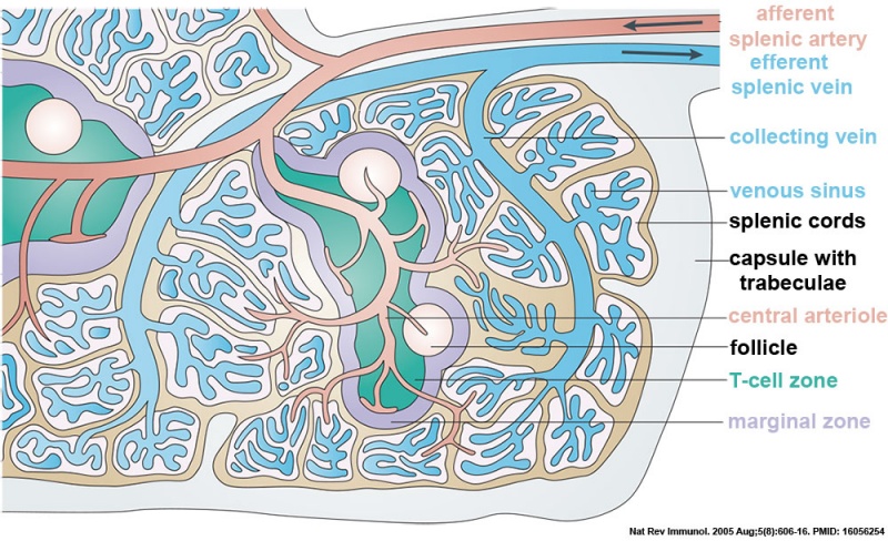
Spleen Structure
This cartoon shows the main structural components of the spleen.
- afferent splenic artery branches into central arterioles, which are sheathed by white-pulp areas.
- white pulp areas consist of the T-cell zone (also known as the periarteriolar lymphoid sheath, PALS), arterioles and B-cell follicles.
- arterioles end in cords in the red pulp, from where the blood runs into venous sinuses that collect into the efferent splenic vein.
- larger arteries and veins run together in connective-tissue trabeculae, which are continuous with the capsule that surrounds the spleen.
- Links: spleen
Reference
Mebius RE & Kraal G. (2005). Structure and function of the spleen. Nat. Rev. Immunol. , 5, 606-16. PMID: 16056254 DOI.
Copyright
Reprinted by permission from Macmillan Publishers Ltd: [Nat Rev Immunol.] (Nature Reviews Immunology 5, 606-616 (August 2005)) | doi:10.1038/nri1669), copyright (2005)
Fig. 1. cartoon relabelled with colour coded labels and reference.
Cite this page: Hill, M.A. (2024, June 5) Embryology Spleen structure cartoon 01.jpg. Retrieved from https://embryology.med.unsw.edu.au/embryology/index.php/File:Spleen_structure_cartoon_01.jpg
- © Dr Mark Hill 2024, UNSW Embryology ISBN: 978 0 7334 2609 4 - UNSW CRICOS Provider Code No. 00098G
File history
Click on a date/time to view the file as it appeared at that time.
| Date/Time | Thumbnail | Dimensions | User | Comment | |
|---|---|---|---|---|---|
| current | 13:21, 26 February 2016 | | 1,000 × 610 (217 KB) | Z8600021 (talk | contribs) | text recoded to cartoon colour |
| 12:20, 26 February 2016 |  | 1,000 × 610 (213 KB) | Z8600021 (talk | contribs) |
You cannot overwrite this file.
File usage
The following 5 pages use this file: