File:Keibel Mall 2 555.jpg

Original file (1,280 × 904 pixels, file size: 195 KB, MIME type: image/jpeg)
Fig. 555. Median section of a human embryo of 50 mm head-foot length. (Embryo R. Meyer 257, from the collection of Professor R. Meyer, Berlin; slide 16, row 3, section 1.) X 8. On the development of the genital cord the first indication of a sexual difference appears. In the male the two mesonephric folds fuse with the posterior wall of the bladder before they unite to form the genital cord, and this fusion persists also after the union. The urogenital sinus is cut lengthwise; the anterior border of its external orifice reaches already the coronary sulcus of the glans. Between the external orifice of tlfc sinus and the anus the aboral periphery of the phallus and the perineum are beginning to form.
File history
Yi efo/eka'e gwa ebo wo le nyangagi wuncin ye kamina wunga tinya nan
| Gwalagizhi | Nyangagi | Dimensions | User | Comment | |
|---|---|---|---|---|---|
| current | 09:23, 15 November 2018 | | 1,280 × 904 (195 KB) | Z8600021 (talk | contribs) | |
| 09:22, 15 November 2018 | 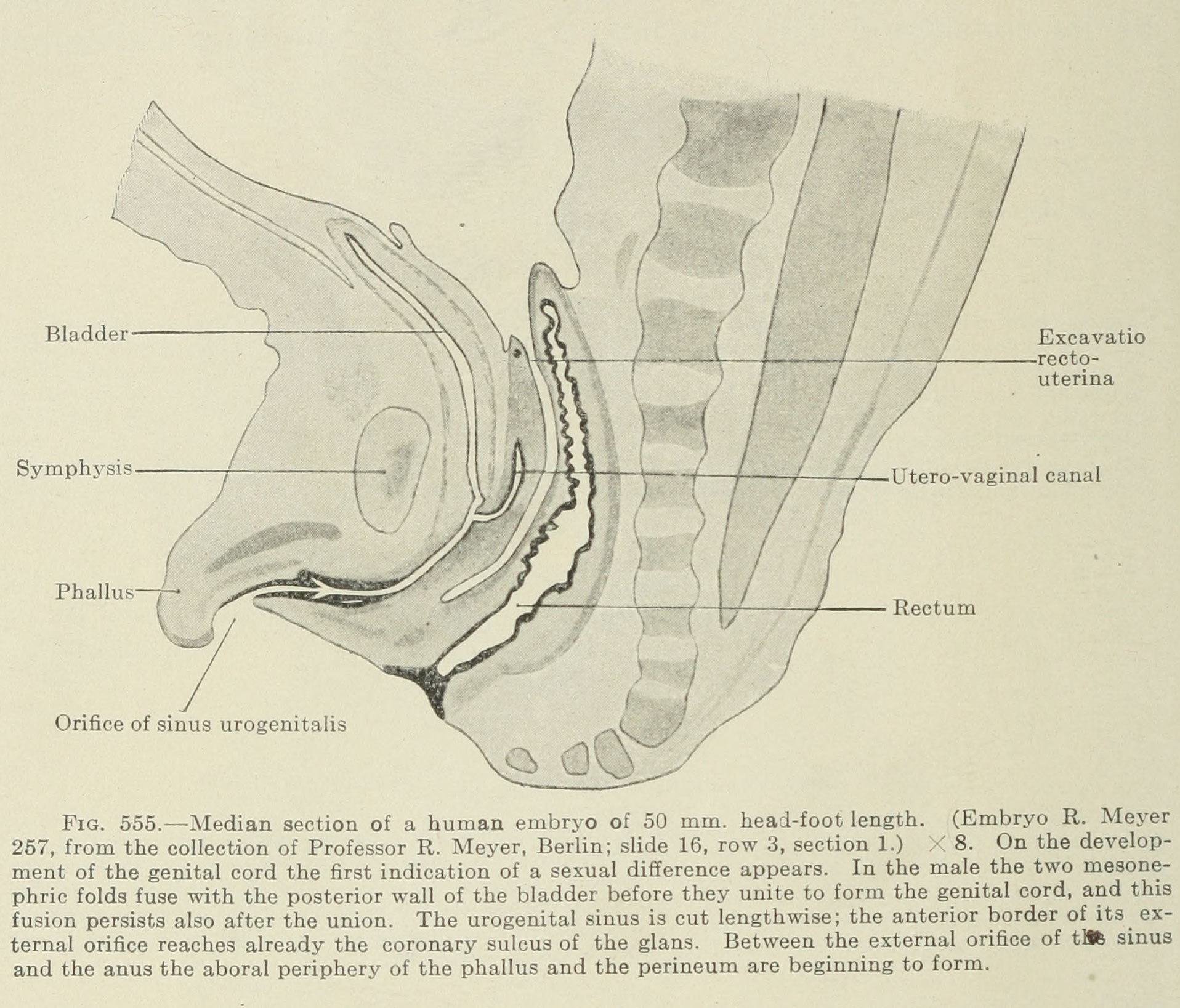 | 1,916 × 1,637 (437 KB) | Z8600021 (talk | contribs) | Fig. 555. Median section of a human embryo of 50 mm head-foot length. (Embryo R. Meyer 257, from the collection of Professor R. Meyer, Berlin; slide 16, row 3, section 1.) X 8. On the development of the genital cord the first indication of a sexual dif... |
You cannot overwrite this file.
File usage
The following 2 pages use this file:


