File:Keibel Mall 2 616.jpg

Original file (1,280 × 1,301 pixels, file size: 309 KB, MIME type: image/jpeg)
Fig. 616 a and b. Transverse section through the testis of a human embryo of the 7th month. (Embryo obtained by operation. From the collection of the Anatomical Institute, Zurich.) The testis cords have a rounded form; their cells are partly indifferent epithelial cells that are beginning to arrange themselves like an epithelium, and partly genital cells, scattered as spermatogonia among the indifferent cells; in the centre is the remains of an intermediate cord with Leydig cells. The tunica albuginea is already formed of a distinctly fibrous connective tissue.
File history
Yi efo/eka'e gwa ebo wo le nyangagi wuncin ye kamina wunga tinya nan
| Gwalagizhi | Nyangagi | Dimensions | User | Comment | |
|---|---|---|---|---|---|
| current | 13:29, 12 November 2018 | | 1,280 × 1,301 (309 KB) | Z8600021 (talk | contribs) | |
| 13:27, 12 November 2018 | 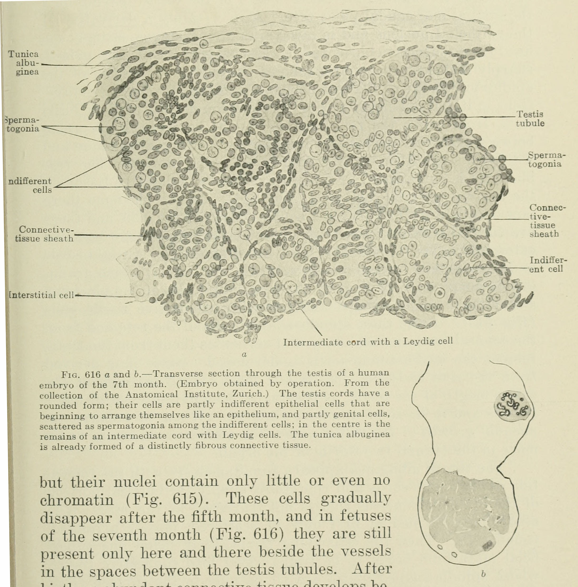 | 2,191 × 2,227 (687 KB) | Z8600021 (talk | contribs) | Fig. 616 a and b. Transverse section through the testis of a human embryo of the 7th month. (Embryo obtained by operation. From the collection of the Anatomical Institute, Zurich.) The testis cords have a rounded form; their cells are partly indifferen... |
You cannot overwrite this file.
File usage
The following 2 pages use this file:


