File:Cullen1916 fig33.jpg

Original file (1,154 × 2,052 pixels, file size: 422 KB, MIME type: image/jpeg)
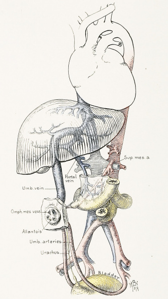
Fig. 33. A Diagrammatic Representation of the Umbilical Region of a Fetus at Term
An exceptional feature of the picture is the persistence of the omphalomesenteric structures in the form of a welldefined vascular Meckel's diverticulum. Note the relationship between the omphalomesenteric vessels, the superior mesenteric artery, and the portal vein. In the cord the vitelline vessels still persist in the organized exoccelom. At birth all trace of the omphalomesenteric structure has usually disappeared, but the allantoic cord often persists, and is seen between and below the umbilical arteries. It is continuous with the urachus, which may have a number of spindleshaped dilatations down to the bladder. These may or may not have lumina.
File history
Yi efo/eka'e gwa ebo wo le nyangagi wuncin ye kamina wunga tinya nan
| Gwalagizhi | Nyangagi | Dimensions | User | Comment | |
|---|---|---|---|---|---|
| current | 13:46, 28 October 2018 | | 1,154 × 2,052 (422 KB) | Z8600021 (talk | contribs) | |
| 13:41, 28 October 2018 | 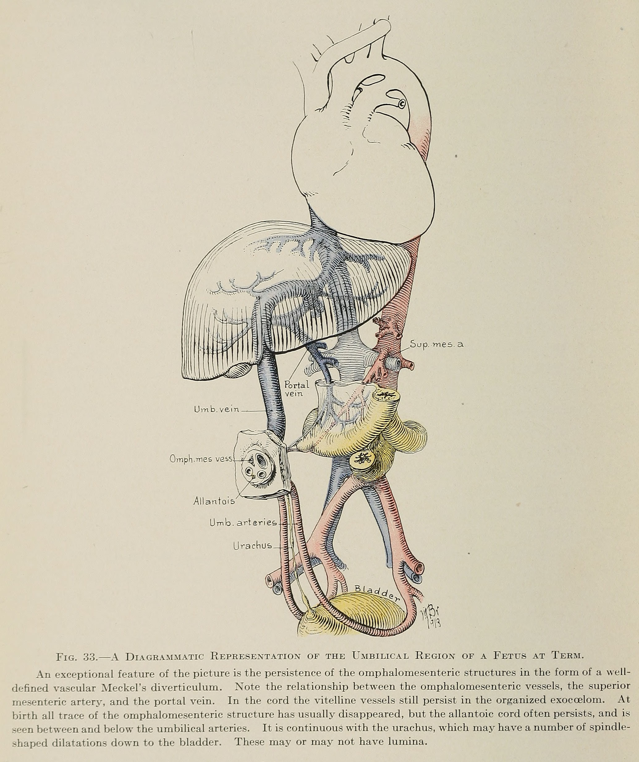 | 2,068 × 2,466 (690 KB) | Z8600021 (talk | contribs) | ==Fig. 33. A Diagrammatic Representation of the Umbilical Region of a Fetus at Term== An exceptional feature of the picture is the persistence of the omphalomesenteric structures in the form of a welldefined vascular Meckel's diverticulum. Note the re... |
You cannot overwrite this file.
File usage
The following 3 pages use this file: