File:Cullen1916 fig09.jpg
Original file (1,280 × 1,683 pixels, file size: 459 KB, MIME type: image/jpeg)
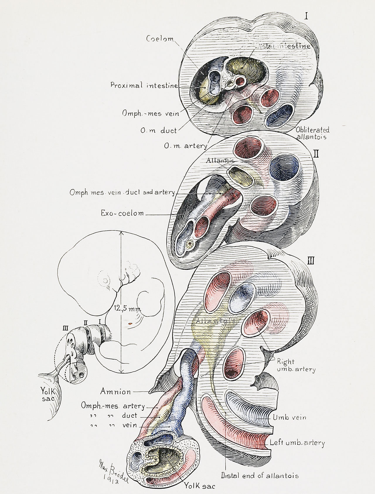
Fig. 9. Graphic Reconstruction of the Umbilical Cord of a Human Embryo 12.5 mm in Length
(Mall collection, No. 317)
The figure to the left shows the manner in which the cord has been cut into the portions I, II, and III. On the right are shown frontal views of the three sections. They are represented as being seniitransparent, in order to give a clear idea of the course of the contained structures.
Section I: Embryonic end of cord, showing ccelomic ring through which protrudes the primitive intestine. Note
the position of the proximal intestine (jejunum) and the distal intestine (ileum). To the right and above, the
omphalomesenteric vessels are seen emerging by the side of the intestine. The vein comes from above the gut and
the artery from below; between them lies the duct, which is still connected with the intestine. A delicate mesenteriolum accompanies these structures for a short distance into the cord (cf. also Fig. 7). The umbilical arteries and
vein are seen in the lower part of the cord. Here the allantois lies between the arteries and becomes obliterated.
Section II: This section of the cord is farther from the umbilicus, and shows a twist of the structures, somewhat
after the manner of the left turn of a screw. The exoccelom with its contents now lies to the right ; the umbilical vessels
and allantois have shifted to the left. The allantois has again increased in size, and lies between the umbilical arteries
and the exoccelom.
Section III: This section of the cord is still farther from the umbilicus. It shows the amniotic ring, through which
emerge the omphalomesenteric duct and its vessels. The allantois decreases rapidly in size and becomes lost between
the left umbilical artery and the umbilical vein.
| Historic Disclaimer - information about historic embryology pages |
|---|
 |
- Figure Links: 1 Human embryo 0.7 mm | 2 Human embryo 1.7 mm | 3 Human embryo 2.5 mm | 4 Human embryo 3.5 mm | 5 Human embryo 5 mm | 6 Human embryo 7 mm | 7 Human embryo 7 mm | 8 Human embryo 10 mm | 9 Human embryo 12.5 mm | 10 Human embryo 10 mm | 11 Human embryo 23 mm | 12 Human embryo 3 cm | 13 Human embryo 4.5 cm sagittal | 14 Human Embryo 4.5 cm | 15 Human Embryo 5.2 cm | 16 Human Embryo 6.5 cm | 17 Human Embryo 7.5 cm | 18 Human Embryo 9 cm | 19 Human Embryo 10 cm | 20 Human Embryo 12 cm | 21 Human Embryo 12 cm | 22 Human Embryo 12 cm | 23 Human Embryo 12 cm Cord | 28 Fetus Five Months | 30 Ventral Heria | 31 Human Embryo 5.5 cm | 32 Term Human | 33 Term Human | [[Figures
Reference
Cullen TS. Embryology, anatomy, and diseases of the umbilicus together with diseases of the urachus. (1916) W. B. Saunders Company, Philadelphia And London.
Cite this page: Hill, M.A. (2024, June 5) Embryology Cullen1916 fig09.jpg. Retrieved from https://embryology.med.unsw.edu.au/embryology/index.php/File:Cullen1916_fig09.jpg
- © Dr Mark Hill 2024, UNSW Embryology ISBN: 978 0 7334 2609 4 - UNSW CRICOS Provider Code No. 00098G
File history
Click on a date/time to view the file as it appeared at that time.
| Date/Time | Thumbnail | Dimensions | User | Comment | |
|---|---|---|---|---|---|
| current | 17:04, 27 October 2018 | | 1,280 × 1,683 (459 KB) | Z8600021 (talk | contribs) | |
| 17:03, 27 October 2018 |  | 2,077 × 3,333 (1.2 MB) | Z8600021 (talk | contribs) | Fig. 9. Graphic Reconstruction of the Umbilical Cord of a Human Embryo 12.5 mm. in Length. (Mall collection, No. {{CE317}}.) The figure to the left shows the manner in which the cord has been cut into the portions I, II, and III. On the right are sh... |
You cannot overwrite this file.
File usage
The following 3 pages use this file: