File:Cullen1916 fig01.jpg
Original file (1,280 × 929 pixels, file size: 354 KB, MIME type: image/jpeg)
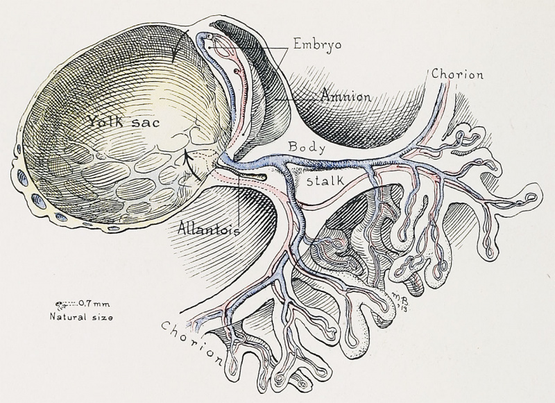
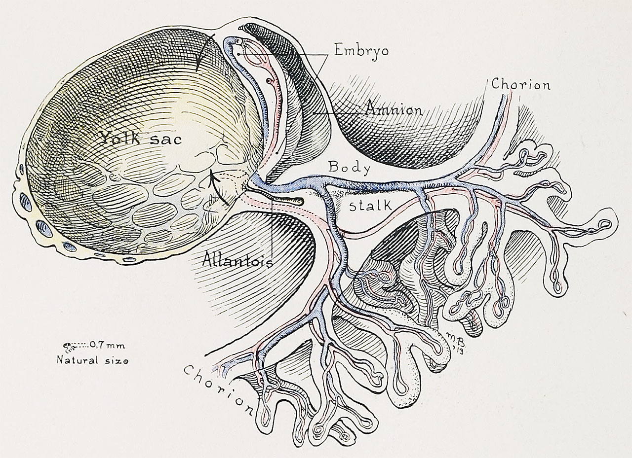
Fig. 1. Sagittal Section Showing a Very Early Stage in the Formation of the Umbilicus and Allantois
(Human embryo, 0.7 mm. long.)
Note the origin of the allantois from the cavity of the yolk-sac. The umbilical region will be formed by a gradua
approximation of the cranial and caudal ends of the yolk-sac, as indicated by the arrows. The embryonic surface of
the yolk-sac will later become the alimentary canal.
Reference
Cullen TS. Embryology, anatomy, and diseases of the umbilicus together with diseases of the urachus. (1916) W. B. Saunders Company, Philadelphia And London.
Cite this page: Hill, M.A. (2026, October 3) Embryology Cullen1916 fig01.jpg. Retrieved from https://embryology.med.unsw.edu.au/embryology/index.php/File:Cullen1916_fig01.jpg
- © Dr Mark Hill 2026, UNSW Embryology ISBN: 978 0 7334 2609 4 - UNSW CRICOS Provider Code No. 00098G
File history
Yi efo/eka'e gwa ebo wo le nyangagi wuncin ye kamina wunga tinya nan
| Gwalagizhi | Nyangagi | Dimensions | User | Comment | |
|---|---|---|---|---|---|
| current | 16:57, 27 October 2018 | | 1,280 × 929 (354 KB) | Z8600021 (talk | contribs) | |
| 16:56, 27 October 2018 | 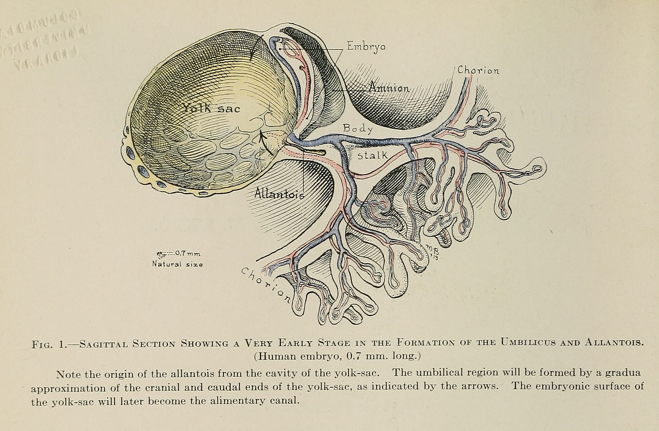 | 2,163 × 1,417 (587 KB) | Z8600021 (talk | contribs) | Fig. 1. — Sagittal Section Showing a Very Early Stage in the Formation of the Umbilicus and Allantois. (Human embryo, 0.7 mm. long.) Note the origin of the allantois from the cavity of the yolk-sac. The umbilical region will be formed by a gradua a... |
You cannot overwrite this file.
File usage
The following 3 pages use this file: