File:Hair Follicle.jpg
From Embryology
Size of this preview: 643 × 599 pixels. Other resolutions: 2,197 × 2,048 pixels | 2,622 × 2,444 pixels.
Original file (2,622 × 2,444 pixels, file size: 2.14 MB, MIME type: image/jpeg)
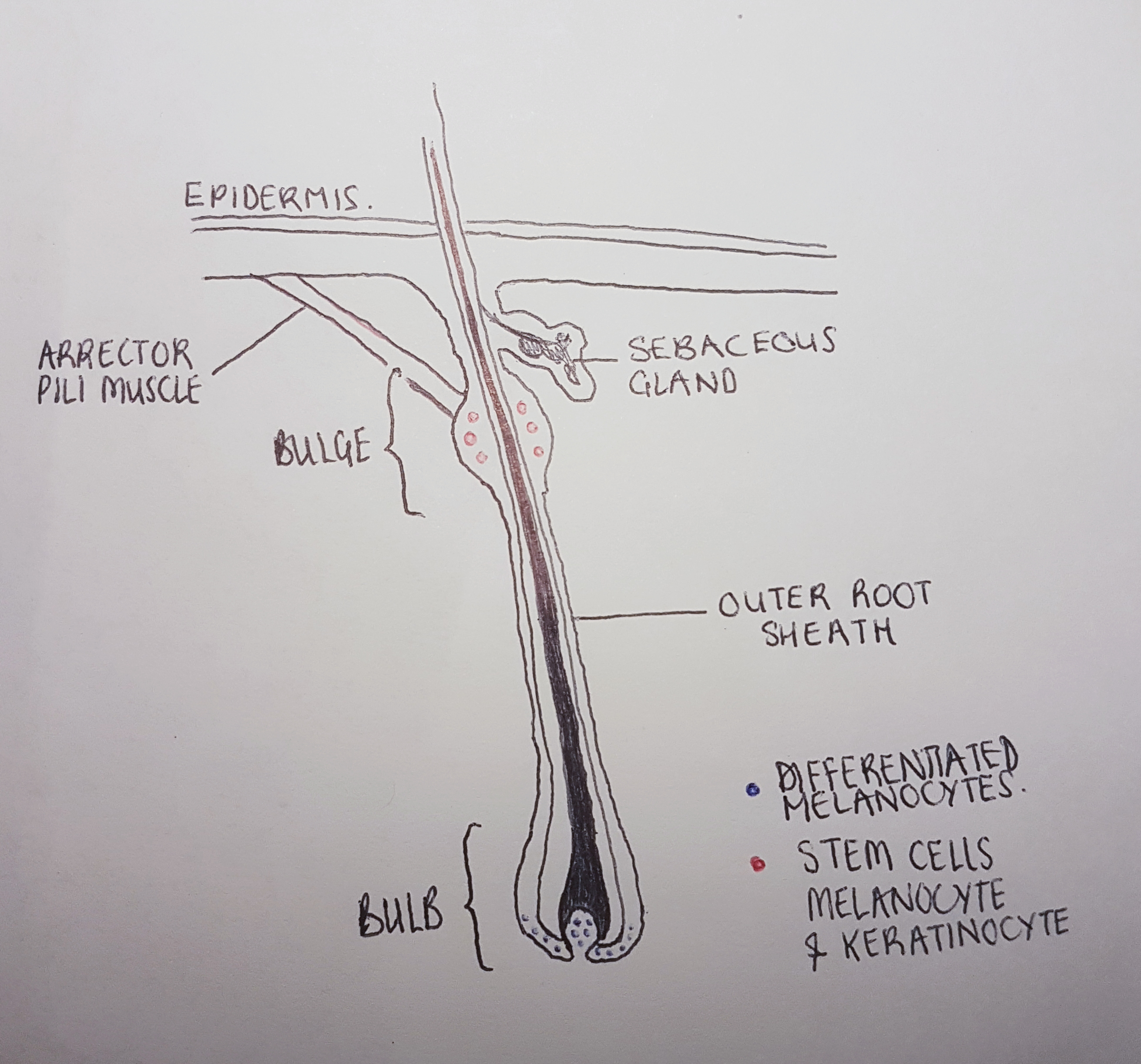
Diagram of the hair follicle at anagen phase: populations of melanocyte and keratinocyte stem cells are located in the hair bulge in red. Differentiated melanocytes in the hair bulb are in blue. These supply melanosomes for hair pigmentation to the keratinocytes that form the hair shaft.
Drawn by student - gathered information from diagrams of various sources.
- Note - This image was originally uploaded as part of an undergraduate science student 2018 project and may contain inaccuracies in either description or acknowledgements. Students have been advised in writing concerning the reuse of content and may accidentally have misunderstood the original terms of use. If image reuse on this non-commercial educational site infringes your existing copyright, please contact the site editor for immediate removal.
File history
Click on a date/time to view the file as it appeared at that time.
| Date/Time | Thumbnail | Dimensions | User | Comment | |
|---|---|---|---|---|---|
| current | 21:48, 16 October 2018 | | 2,622 × 2,444 (2.14 MB) | Z5229132 (talk | contribs) | Diagram of the hair follicle at anagen phase: populations of melanocyte and keratinocyte stem cells are located in the hair bulge in red. Differentiated melanocytes in the hair bulb are in blue. These supply melanosomes for hair pigmentation to the ke... |
You cannot overwrite this file.
File usage
The following 2 pages use this file: