File:Cardiac melanocyte in an embryonic mouse pup.png
From Embryology
Size of this preview: 800 × 557 pixels. Other resolution: 1,424 × 992 pixels.
Original file (1,424 × 992 pixels, file size: 427 KB, MIME type: image/png)

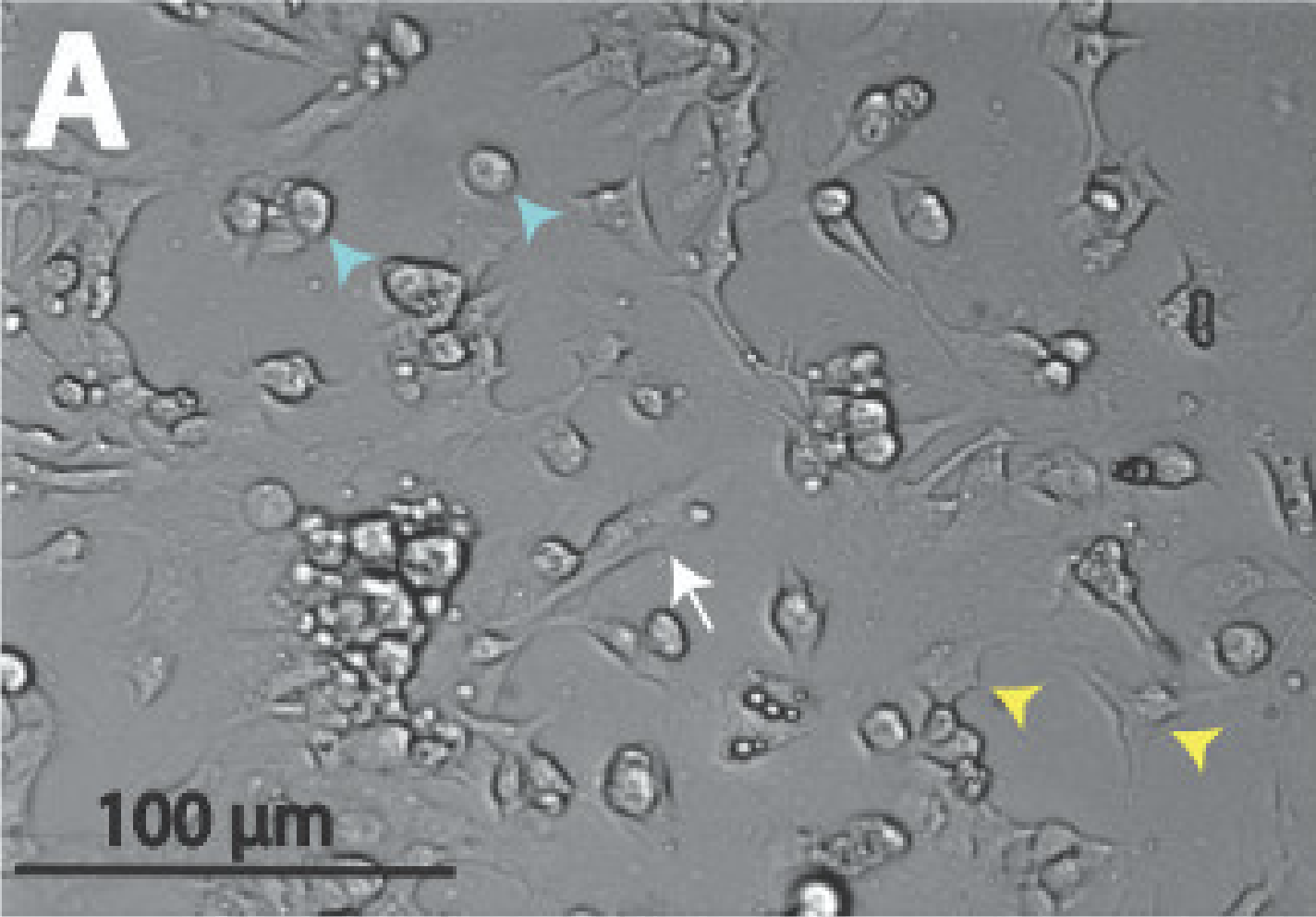
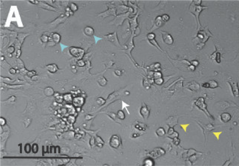
Figure 3.
A differential interference contrast (DIC) microscopical image of a cardiac melanocyte present in an isolated atrial cell of an embryonic mouse pup. The white arrow indicates the cardiac melanocyte that is found amidst the atrial myocytes that are marked by the yellow arrowheads.
Reference
Hwang H, Liu F, Levin MD & Patel VV. (2014). Isolating primary melanocyte-like cells from the mouse heart. J Vis Exp , , 4357. PMID: 25285608 DOI.
Copyright
© 2014, Journal of Visualized Experiments
- Note - This image was originally uploaded as part of an undergraduate science student project and may contain inaccuracies in either description or acknowledgements. Students have been advised in writing concerning the reuse of content and may accidentally have misunderstood the original terms of use. If image reuse on this non-commercial educational site infringes your existing copyright, please contact the site editor for immediate removal.
File history
Yi efo/eka'e gwa ebo wo le nyangagi wuncin ye kamina wunga tinya nan
| Gwalagizhi | Nyangagi | Dimensions | User | Comment | |
|---|---|---|---|---|---|
| current | 23:10, 28 August 2018 | | 1,424 × 992 (427 KB) | Z5164785 (talk | contribs) | 400px Figure 3. A differential interference contrast (DIC) microscopical image of a cardiac melanocyte present in an isolated atrial cell of an embryonic mouse pup. The white arrow indicates... |
You cannot overwrite this file.
File usage
The following 3 pages use this file: