File:Hindbrain neural crest migration.jpg
Hindbrain_neural_crest_migration.jpg (450 × 545 pixels, file size: 48 KB, MIME type: image/jpeg)
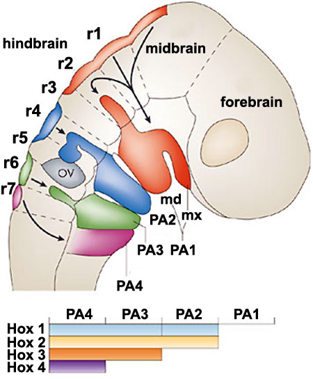
Hindbrain neural crest migration
A schematic diagram of a chick head at embryonic day two, showing pathways of neural crest migration in the chick and mouse embryo and patterns of Hox gene expression in the branchial arches (BAs)42, 102, 169, 170. FB, forebrain; HB, hindbrain; MB, midbrain; Md, mandibular part of BA1; Mx, maxillary part of BA1; OV, otic vesicle; r, rhombomere.
Original Figure: 4 http://www.nature.com/nrn/journal/v8/n11/fig_tab/nrn2254_F4.html
Reference
<pubmed>17948031</pubmed>
Adapted by permission from Macmillan Publishers Ltd: Nature Reviews Neuroscience (<pubmed>17948031</pubmed>), copyright (2007)
File history
Yi efo/eka'e gwa ebo wo le nyangagi wuncin ye kamina wunga tinya nan
| Gwalagizhi | Nyangagi | Dimensions | User | Comment | |
|---|---|---|---|---|---|
| current | 16:23, 31 August 2010 | | 450 × 545 (48 KB) | S8600021 (talk | contribs) | ==Hindbrain neural crest migration== A schematic diagram of a chick head at embryonic day two, showing pathways of neural crest migration in the chick and mouse embryo and patterns of Hox gene expression in the branchial arches (BAs)42, 102, 169, 170. FB |
You cannot overwrite this file.