File:Shanklin1940 fig03.jpg
From Embryology

Size of this preview: 376 × 599 pixels. Other resolution: 636 × 1,013 pixels.
Original file (636 × 1,013 pixels, file size: 96 KB, MIME type: image/jpeg)
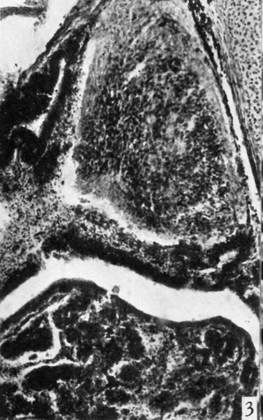
Fig. 3. Photomicrograph showing from above downward the pars nervosa
The pars intermedia, cleft, and the pars anterior of a 3-month human foetus. x 95.
Reference
Shanklin WM. Differentiation of pituicytes in the human foetus. (1940) J Anat. 74(4): 459-63. PMID 17104829
Cite this page: Hill, M.A. (2024, June 5) Embryology Shanklin1940 fig03.jpg. Retrieved from https://embryology.med.unsw.edu.au/embryology/index.php/File:Shanklin1940_fig03.jpg
- © Dr Mark Hill 2024, UNSW Embryology ISBN: 978 0 7334 2609 4 - UNSW CRICOS Provider Code No. 00098G
File history
Click on a date/time to view the file as it appeared at that time.
| Date/Time | Thumbnail | Dimensions | User | Comment | |
|---|---|---|---|---|---|
| current | 21:19, 26 June 2018 | | 636 × 1,013 (96 KB) | Z8600021 (talk | contribs) | ==Fig. 3. Photomicrograph showing from above downward the pars nervosa== The pars intermedia, cleft, and the pars anterior of a 3-month human foetus. x 95. ===Reference=== {{Ref-Shanklin1940}} {{Footer}} |
You cannot overwrite this file.
File usage
The following page uses this file: