File:Shanklin1940 plate02.jpg
Original file (1,411 × 2,131 pixels, file size: 210 KB, MIME type: image/jpeg)
Plate 2
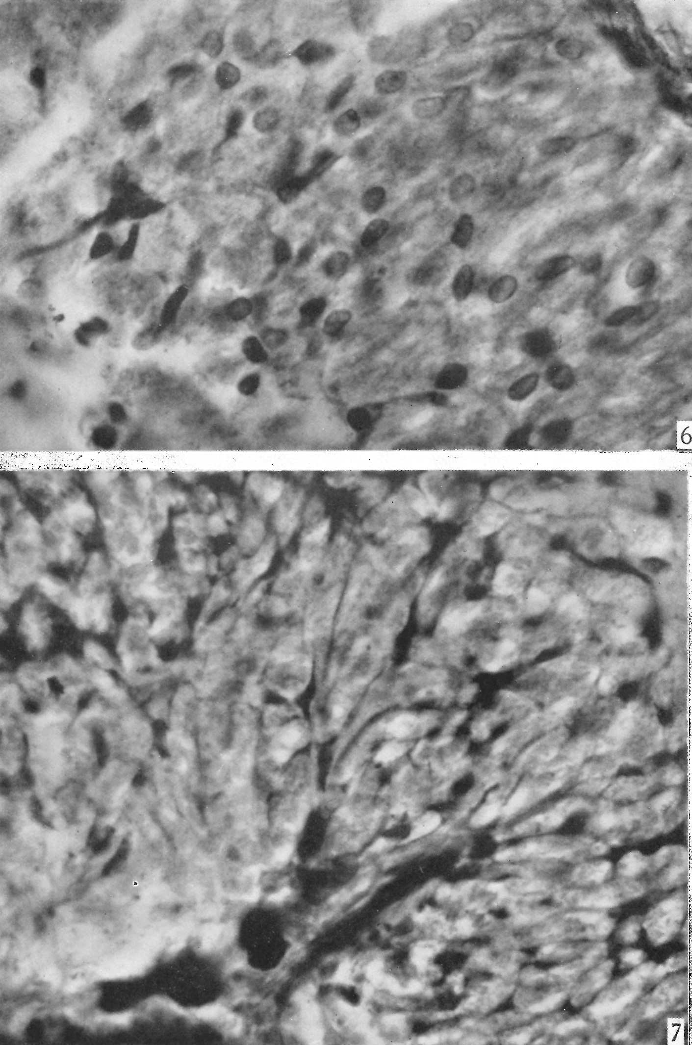
Fig. 6. Photomicrograph of same section shown in P1. I, fig. 4 showing details of cells and intercellular granules in the zone distal to the pars intermedia. x 725.
Fig. 7. Photomicrograph demonstrating details of the proximal area of the same section shown in P1. I, fig. 4. Note the prominent trabecula, the accompanying blood-vessels stained black, the - processes attached to the septum, and processes anastomosing with one another to form a reticulum. Due to lack of focal depth the reticulum is not adequately represented. x 725.
Reference
Shanklin WM. Differentiation of pituicytes in the human foetus. (1940) J Anat. 74(4): 459-63. PMID 17104829
Cite this page: Hill, M.A. (2024, June 5) Embryology Shanklin1940 plate02.jpg. Retrieved from https://embryology.med.unsw.edu.au/embryology/index.php/File:Shanklin1940_plate02.jpg
- © Dr Mark Hill 2024, UNSW Embryology ISBN: 978 0 7334 2609 4 - UNSW CRICOS Provider Code No. 00098G
File history
Click on a date/time to view the file as it appeared at that time.
| Date/Time | Thumbnail | Dimensions | User | Comment | |
|---|---|---|---|---|---|
| current | 21:11, 26 June 2018 | | 1,411 × 2,131 (210 KB) | Z8600021 (talk | contribs) | |
| 21:10, 26 June 2018 | 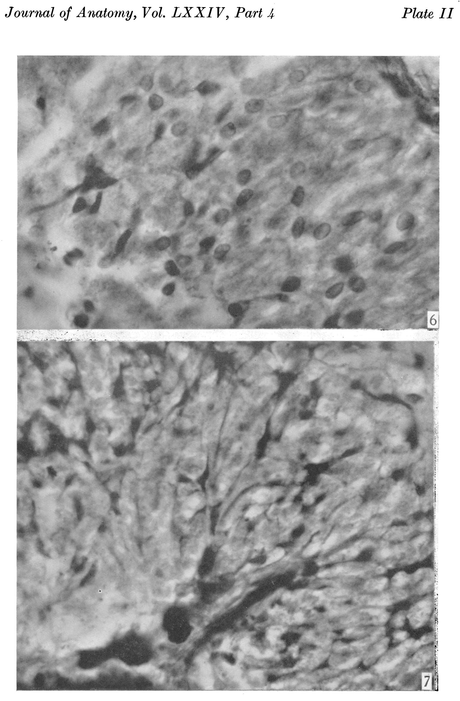 | 1,551 × 2,362 (368 KB) | Z8600021 (talk | contribs) |
You cannot overwrite this file.
File usage
The following page uses this file: